Undifferentiated shock
Bottom line
- Hypotension is not required for shock. Look for tachycardia and poor perfusion: abnormal skin, low urine output and altered mentation. Measure lactate in every patient with suspected shock.1,2,3
- Name the type: hypovolemic, cardiogenic, obstructive or distributive. Types can overlap. Echocardiography is the suggested first-line tool to tell them apart.1,3
- Ultrasound sharpens the diagnosis, but has not shown a survival benefit. Immediate scanning put the right diagnosis first in 80% vs 50% of patients. In SHoC-ED, 30-day survival did not differ.4,5
- Before giving more fluid after the first bolus, check whether the patient will respond. A fluid challenge is 200–500 mL over 5–10 minutes, with reassessment.1,2
- Norepinephrine, not dopamine. In SOAP II, dopamine caused about twice the arrhythmias (24.1% vs 12.4%). Death rates were higher with dopamine in the cardiogenic shock subgroup. Start with a MAP of 65 or more, then individualize.2,3,6
First steps
- Monitor and examine often: heart rate, blood pressure, temperature, skin perfusion, urine output and mental status.3
- Check perfusion: time the capillary refill, and note skin temperature and mottling.1,2 Send a lactate.3
- Look at the context: trauma, infection, chest pain. The skin and the neck veins often point to the type.3
- Scan the heart early when the exam doesn't make the type clear.1,3
- Treat the cause and support the circulation together: fluids, and vasopressors if needed, with frequent reassessment.3
- If shock doesn't respond, or a vasopressor is running, place an arterial line and plan cardiac output or stroke volume monitoring.1,2
Recognize and classify
Shock is acute circulatory failure in which cells can't use enough oxygen. Lactate rises as a result.3 The usual definition of hypotension is a systolic pressure under 90 mm Hg, a MAP under 65, or a drop of 40 mm Hg or more from baseline. It is common but shouldn't be required for the diagnosis.3 Compensatory vasoconstriction can hold the pressure up while perfusion is already falling.3
| Type | Mechanism and examples | Hemodynamic pattern | ED share* |
|---|---|---|---|
| Hypovolemic | Lost volume, internal or external (bleeding, fluid loss) | Low cardiac output; low pressures and volumes | 30.8% |
| Cardiogenic | Pump failure: ischemia, infarction, myopathy, myocarditis, major arrhythmia | Low cardiac output; raised pressures and volumes | 14.0% |
| Obstructive | Pulmonary embolism, tension pneumothorax, cardiac tamponade | Low cardiac output; high pulmonary pressure and a dilated right heart. Tamponade compresses all chambers. | 0.9% |
| Distributive | Lost vascular tone: sepsis, anaphylaxis, spinal injury | Usually high cardiac output | 27.2% septic, 23.4% non-septic |
Types and patterns from the ESICM consensus.3 *Shares are from 1,553 Danish ED patients with a systolic pressure of 100 mm Hg or less plus at least one organ failure. Another 3.7% had other conditions.7 In ICU patients from SOAP II, septic shock made up 62%, cardiogenic 17% and hypovolemic 16%.3 More than one type can coexist.3
The type predicts the outcome. In the Danish cohort, 7-day mortality ranged from 12.7% to 34.6% by type. Septic and cardiogenic shock independently predicted death within 7 days (HR 1.46 and 2.15).7
Shock index (heart rate ÷ systolic pressure)
From 111,019 first-time ED visits, with an overall 30-day mortality of 3.0%. The link was weaker in patients 65 or older (OR 8.2 vs 18.9 for younger patients), on beta- or calcium channel blockers (6.4 vs 12.3), or with hypertension (8.0 vs 12.9). A shock index of 1 or more still signaled high risk in every group.8
Bedside ultrasound
- ESICM suggests echocardiography as the first-line imaging test for the type of shock (a graded statement), and says it changes management (good practice).1,2 The 2014 consensus noted that a basic look usually takes under 2 minutes, even for physicians with little training.3
- Pooled accuracy (12 studies, 1,132 patients): sensitivity 0.78–0.90 and specificity 0.92–0.98 across the four types. The positive likelihood ratio was highest for obstructive shock (40).9
- Faster, better diagnosis: in 184 ED patients with nontraumatic hypotension, the immediate-scan group had a median of 4 possible diagnoses at 15 minutes, vs 9 with delayed scanning. The final diagnosis was ranked most likely in 80% vs 50%.4
- No survival benefit shown: SHoC-ED randomized 273 hypotensive ED patients (systolic under 100 mm Hg or shock index over 1). Survival was 104 of 136 with ultrasound vs 102 of 134 without. Fluid use, CT use and length of stay were also similar. The most common diagnosis was occult sepsis.5
| Type | What the scan shows | Pooled sensitivity | Pooled specificity |
|---|---|---|---|
| Obstructive | Dilated right heart; in tamponade, small, compressed chambers | 0.82 | 0.98 |
| Cardiogenic | Weak contraction; large, high-pressure chambers | 0.78 | 0.96 |
| Hypovolemic | Small, underfilled chambers | 0.90 | 0.92 |
| Distributive | High cardiac output | 0.79 | 0.96 |
Drawn for this page. Patterns from the ESICM consensus; accuracy from a 2023 meta-analysis.3,9 A right-to-left ventricular end-diastolic area ratio of 0.6 to 1 suggests moderate right ventricular dilation, and over 1 suggests severe dilation.3
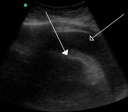
The IVC is a weak guide on its own. In SHoC-ED patients breathing spontaneously, an IVC over 2.5 cm that collapsed less than 50% detected volume overload with a sensitivity of 85.7% and specificity of 86.4%. Only 7 of 125 patients were overloaded.10 ESICM warns against steering fluids by any single static preload number.2,3
ESICM supports echo as the first look at shock type.1,3 SHoC-ED found no survival gain.5 Reviewers note that the trial stopped early (273 of 400 planned) for slow recruitment and futility. They also note that no change in management was recorded after the scans, and that a diagnostic test may be a poor fit for a mortality endpoint.11
Lactate and capillary refill
- Lactate: measure it whenever you suspect shock. It is usually above 2 mmol/L (2 mEq/L) in shock. Repeat it to guide treatment: about every 2 hours for the first 8 hours, then every 8–12 hours.3
- Lactate beats blood pressure for prognosis, but it can rise for reasons other than poor perfusion.3
- Capillary refill time is ESICM's first skin-perfusion check, with skin temperature and mottling as add-ons.1,2
- After a fluid bolus, judge the effect on capillary refill, mottling, venous oxygen saturation, CO2-derived values and lactate.2
What the trials show (septic shock only)
- ANDROMEDA-SHOCK (n = 424, 28 ICUs): an 8-hour protocol targeting capillary refill vs lactate. 28-day mortality was 34.9% vs 43.4% (HR 0.75, 95% CI 0.55–1.02), not significant. Organ dysfunction at 72 hours was lower (mean SOFA 5.6 vs 6.6).12
- ANDROMEDA-SHOCK-2 (n = 1,467, 86 centers): a personalized protocol targeting capillary refill, including fluid responsiveness and bedside echo, beat usual care on a composite outcome (win ratio 1.16, 95% CI 1.02–1.33). The gain came mostly from a shorter duration of vital support.13
Fluids, vasopressors and inotropes
Fluids
- Too little and too much fluid both harm. Start fluids at once when preload is obviously very low, as in overt bleeding.3
- After the first fluids, check fluid responsiveness before giving more, and weigh the expected benefit against the risk.1,2
- Use dynamic tests over static values such as CVP. Don't target a set CVP.1,2,3 ESICM strongly recommends passive leg raising in ventilated patients, with or without spontaneous breaths (high certainty).2
Blood pressure targets
| Situation | Target |
|---|---|
| Most shock | Start at MAP ≥ 65 mm Hg, then individualize2,3 |
| Cardiogenic shock | An initial MAP ≥ 65 may be considered2 |
| Septic shock with chronic hypertension, or high CVP | A higher MAP may be considered if the patient improves with it2 |
| Trauma, uncontrolled bleeding, no brain injury | Systolic 80–90 mm Hg (MAP 50–60) until major bleeding is stopped2 |
| Traumatic brain injury with GCS ≤ 8 | MAP ≥ 80 mm Hg2 |
Drugs
| Agent | Adult dose | Notes |
|---|---|---|
| Vasopressors | ||
| Norepinephrine | Start 8–12 mcg/min, then titrate. Average maintenance 2–4 mcg/min. | Fewer arrhythmias than dopamine in SOAP II. The label says to correct hypovolemia first.6,14 |
| Epinephrine | 0.05–2 mcg/kg/min, titrated to MAP | Labeled for hypotension in septic shock.15 |
| Vasopressin | Septic shock: start 0.01 units/min; range 0.01–0.07 units/min | Titrate by 0.005 units/min every 10–15 min.16 |
| Dopamine | Not first line | More arrhythmias than norepinephrine. Higher mortality in cardiogenic shock.6 |
| Inotrope | ||
| Dobutamine | Start 0.5–1 mcg/kg/min; usual range 2–20 mcg/kg/min | Only for poor cardiac function with low output and hypoperfusion after preload is optimized.3,17 |
| Extravasation | ||
| Phentolamine | 5–10 mg in 10–15 mL normal saline, infiltrated into the area | Infiltrate as soon as possible after norepinephrine extravasation.14 |
Unreviewed doses. Verify against the current label and your institution's protocol before giving anything.
Doses from the FDA labels. Follow institutional protocols for concentrations and titration.
- SOAP II (n = 1,679 with shock): 28-day mortality was 52.5% with dopamine vs 48.5% with norepinephrine, not significant. Arrhythmias were 24.1% vs 12.4%. Among 280 patients with cardiogenic shock, dopamine was linked to more deaths.6
- Don't give inotropes for a weak heart alone. Add them only when low output and hypoperfusion persist after preload is optimized.3
Peripheral vasopressors
- In 7 studies (1,382 patients), extravasation occurred in 3.4%. No tissue necrosis or limb ischemia was reported, and all events were managed conservatively or with vasodilators.18
- In 9 studies (1,835 patients), 7% had complications and 96% of those were minor. Complications were less common where studies used a safety protocol (2.9% vs 12%).19
The norepinephrine and epinephrine labels say to infuse into a large vein.14,15 Reviews of peripheral infusion found extravasation uncommon and rarely serious, mostly with short infusions under close watch.18,19 The 2014 ESICM consensus said central access may be preferred once ICU patients need vasopressor infusions.3 For septic shock, the 2026 Surviving Sepsis Campaign suggests starting vasopressors peripherally rather than waiting for central access (conditional, very low certainty).20
Disposition
- Shock that doesn't respond to initial therapy, or needs a vasopressor, calls for an arterial line, cardiac output or stroke volume monitoring, and serial echo.1,2 Admit to a setting that can provide them.
- Patients who respond quickly don't need routine cardiac output measurement.3
- Expect high early mortality: 7-day mortality reached 34.6% in cardiogenic shock and 30.3% in septic shock in ED patients.7
Pitfalls
- Waiting for hypotension. Pressure can hold up while perfusion falls.3
- Trusting a normal shock index in older patients or those on beta- or calcium channel blockers. Its signal is weaker there.8
- Relying on one number, such as a single CVP, IVC size or lactate.3,10
- Giving fluid after fluid without checking responsiveness.1,2
- Reaching for dopamine. It caused twice the arrhythmias in SOAP II.6
- Treating the pressure and forgetting the cause: bleeding, tamponade, tension pneumothorax, pulmonary embolism, sepsis.3
How strong is the evidence?
The 2025 ESICM guideline made 50 statements. Where strong evidence was lacking, the panel issued ungraded good practice statements, and most statements in the official summary are of that kind.1,2 It addresses adult ICU patients, so ED use is an extrapolation.1 The best perfusion-target trials enrolled only septic shock.12,13 The ultrasound trials are small, and SHoC-ED stopped early.4,5,11 SOAP II is the largest vasopressor trial across shock types.6 Peripheral vasopressor data come from observational studies.18,19
On-shift teaching
Questions to ask a learner at the bedside. Tap a question to see the answer. Every answer uses facts cited above.
Q1Does a patient need to be hypotensive to be in shock?
Q2Name the four types of shock and the most common in ED patients.
Q3What does a shock index of 1 or more mean?
Q4If ultrasound improves diagnosis, why didn't SHoC-ED show better survival?
Q5Why norepinephrine over dopamine?
Q6What is a fluid challenge, and how do you judge it?
References
- 1Monnet X, Messina A, Greco M, et al. ESICM guidelines on circulatory shock and hemodynamic monitoring 2025. Intensive Care Med. 2025;51(11):1971–2012.
- 2European Society of Intensive Care Medicine. ESICM guidelines on circulatory shock and hemodynamic monitoring 2025: visual abstract and announcement.
- 3Cecconi M, De Backer D, Antonelli M, et al. Consensus on circulatory shock and hemodynamic monitoring. Task force of the European Society of Intensive Care Medicine. Intensive Care Med. 2014;40(12):1795–1815.
- 4Jones AE, Tayal VS, Sullivan DM, et al. Randomized, controlled trial of immediate versus delayed goal-directed ultrasound to identify the cause of nontraumatic hypotension in emergency department patients. Crit Care Med. 2004;32(8):1703–1708.
- 5Atkinson PR, Milne J, Diegelmann L, et al. Does point-of-care ultrasonography improve clinical outcomes in emergency department patients with undifferentiated hypotension? An international randomized controlled trial from the SHoC-ED investigators. Ann Emerg Med. 2018;72(4):478–489.
- 6De Backer D, Biston P, Devriendt J, et al. Comparison of dopamine and norepinephrine in the treatment of shock (SOAP II). N Engl J Med. 2010;362(9):779–789.
- 7Gitz Holler J, Jensen HK, Henriksen DP, et al. Etiology of shock in the emergency department: a 12-year population-based cohort study. Shock. 2019;51(1):60–67.
- 8Kristensen AK, Holler JG, Hallas J, et al. Is shock index a valid predictor of mortality in emergency department patients with hypertension, diabetes, high age, or receipt of β- or calcium channel blockers? Ann Emerg Med. 2016;67(1):106–113.e6.
- 9Yoshida T, Yoshida T, Noma H, et al. Diagnostic accuracy of point-of-care ultrasound for shock: a systematic review and meta-analysis. Crit Care. 2023;27(1):200.
- 10Dunfield R, Ross P, Dutton D, et al. SHoC-IVC: does assessment of the inferior vena cava by point-of-care ultrasound independently predict fluid status in spontaneously breathing patients with undifferentiated hypotension? CJEM. 2023;25(11):902–908.
- 11The Bottom Line. SHoC-ED: point-of-care ultrasound in undifferentiated hypotension (trial summary and critique).
- 12Hernández G, Ospina-Tascón GA, Damiani LP, et al. Effect of a resuscitation strategy targeting peripheral perfusion status vs serum lactate levels on 28-day mortality among patients with septic shock: the ANDROMEDA-SHOCK randomized clinical trial. JAMA. 2019;321(7):654–664.
- 13ANDROMEDA-SHOCK-2 Investigators; Hernandez G, Ospina-Tascón GA, et al. Personalized hemodynamic resuscitation targeting capillary refill time in early septic shock: the ANDROMEDA-SHOCK-2 randomized clinical trial. JAMA. 2025;334(22):1988–1999.
- 14Norepinephrine bitartrate in sodium chloride injection. FDA prescribing information (DailyMed).
- 15Adrenalin (epinephrine in sodium chloride) injection. FDA prescribing information (DailyMed).
- 16Vasopressin injection. FDA prescribing information (DailyMed).
- 17Dobutamine in dextrose injection. FDA prescribing information (DailyMed).
- 18Tian DH, Smyth C, Keijzers G, et al. Safety of peripheral administration of vasopressor medications: a systematic review. Emerg Med Australas. 2020;32(2):220–227.
- 19Tran QK, Mester G, Bzhilyanskaya V, et al. Complication of vasopressor infusion through peripheral venous catheter: a systematic review and meta-analysis. Am J Emerg Med. 2020;38(11):2434–2443.
- 20Society of Critical Care Medicine. Surviving Sepsis Campaign: International Guidelines for Management of Sepsis and Septic Shock 2026 (recommendation summary).
AI-generated draft. Not reviewed by a physician. This page is for education only and may contain errors, including in doses. Do not use it to diagnose, treat or make decisions about any patient. Rely on your own clinical judgment, your institution's protocols and current drug labeling. Reading this page does not create a physician-patient relationship. Full terms