STEMI
Bottom line
- Get and read a 12-lead ECG within 10 minutes. A first ECG that is not diagnostic does not exclude STEMI. Repeat it, and add posterior leads when V1–V3 show ST depression.1
- Primary PCI is the default. The system goal is first medical contact (FMC) to device in 90 minutes or less, or 120 minutes or less when the patient needs transfer.1 Across 23 trials, primary angioplasty lowered short-term death compared with fibrinolysis (7% vs 9%).2
- If PCI will take more than 120 minutes and symptoms began under 12 hours ago, give a fibrinolytic unless it is contraindicated. Then transfer at once for angiography.1
- Every patient gets aspirin, a P2Y12 inhibitor and a parenteral anticoagulant. Use ticagrelor or prasugrel with primary PCI, and clopidogrel with a fibrinolytic.1
- Give oxygen only if saturation is under 90%.1 In 6,629 patients without hypoxemia, routine oxygen did not change 1-year mortality (5.0% vs 5.1%).3
ECG diagnosis
| Leads | New ST elevation at the J point |
|---|---|
| Any 2 contiguous leads other than V2–V3 | ≥ 1 mm |
| V2–V3, men 40 and older | ≥ 2 mm |
| V2–V3, men under 40 | ≥ 2.5 mm |
| V2–V3, women of any age | ≥ 1.5 mm |
STEMI criteria from the 2025 guideline. Pericarditis, left ventricular hypertrophy, LBBB, Brugada syndrome, ventricular pacing, Takotsubo syndrome and early repolarization can all mimic or hide these changes.1
- Repeat the ECG if the first one is not diagnostic, especially when symptoms persist or the patient gets worse. In a US registry, 11% of patients later diagnosed with STEMI had a nondiagnostic first ECG. Of those, 72.4% had a diagnostic ECG within 90 minutes.1
- Posterior leads (V7–V9) for suspected circumflex occlusion, particularly isolated ST depression of 0.5 mm or more in V1–V3.1
- Right-sided leads for inferior STEMI, to look for right ventricular involvement.1 Right ventricular infarction comes with about 40% of inferior STEMIs, and these patients can become severely hypotensive with nitrates.4
- Look at aVL in suspected inferior MI. Reciprocal ST depression in aVL is a sensitive sign. It was present in 91% of subtle inferior occlusions that did not meet STEMI criteria.4
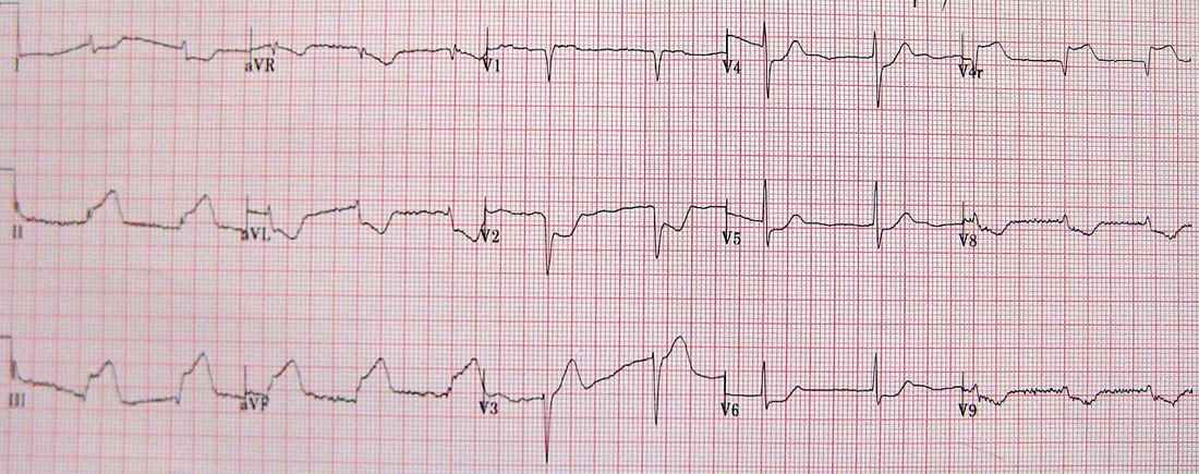
Left bundle branch block
A new or presumed new LBBB is not diagnostic of MI by itself. In a patient without symptoms it is not a STEMI equivalent.1 Look instead for the Smith-modified Sgarbossa criteria. Any one is positive:5
- Concordant ST elevation ≥ 1 mm in at least 1 lead.5
- Concordant ST depression ≥ 1 mm in at least 1 of V1–V3.5
- Discordant ST elevation ≥ 1 mm that is at least 25% of the depth of the preceding S wave (ST/S ratio ≤ −0.25).5,6
In the derivation study (33 occlusions, 129 controls), the modified rule had a sensitivity of 91% and specificity of 90%. The original weighted Sgarbossa rule had 52% and 98%.6 See the LITFL Sgarbossa page for examples.5
The guideline builds its STEMI pathway on millimeter ST-elevation criteria.1 An emergency medicine group argues that these criteria miss many acute coronary occlusions. In a retrospective case-control study of 808 patients, STEMI criteria detected 41% of occlusion MIs, compared with 86% for expert reading of predefined occlusion MI findings. Occlusions missed by STEMI criteria had similar infarct size and mortality, but waited longer for angiography.7 These findings have not been tested in a trial. When the first ECG is not diagnostic and suspicion is high, serial ECGs are recommended.1
First 10 minutes
- ECG within 10 minutes of first contact, read by a trained clinician.1
- Activate the cath lab on a diagnostic ECG. Prehospital activation and advance notice from EMS shorten time to reperfusion.1
- Give aspirin 162–325 mg, chewed, even if the patient already takes aspirin.1
- Don't wait for troponin. Reperfusion should not be delayed for biomarker results.1
- Decide on reperfusion: primary PCI if device time can be 120 minutes or less from first contact. Otherwise a fibrinolytic, if symptoms began under 12 hours ago and there is no contraindication.1
- Treat pain and ischemia: sublingual nitroglycerin if systolic BP is 90 mm Hg or higher. Oxygen only if saturation is under 90%.1
- Look for shock. Do an urgent echo (point-of-care ultrasound is fine) for shock, instability or suspected mechanical complications.1
Reperfusion
| Situation | Recommendation | Class |
|---|---|---|
| PCI-capable hospital | ||
| Symptoms < 12 h | Primary PCI, FMC to device ≤ 90 min (≤ 120 min if transferred) | 1 |
| Symptoms 12–24 h | Primary PCI is reasonable | 2a |
| > 24 h with ongoing ischemia or life-threatening arrhythmia | Primary PCI is reasonable | 2a |
| > 24 h, stable, occluded artery, no ischemia or severe heart failure | No PCI (no benefit) | 3 |
| Non-PCI hospital | ||
| FMC to device expected ≤ 120 min, or fibrinolytic contraindicated | Transfer for primary PCI | 1 |
| Symptoms < 12 h and PCI delay > 120 min | Fibrinolytic, unless contraindicated | 1 |
| Symptoms 12–24 h | Transfer for PCI is reasonable | 2a |
| ST depression only, no suspected posterior STEMI | No fibrinolytic (harm) | 3 |
From the 2025 guideline. Class 1 is a strong recommendation, 2a moderate, 3 no benefit or harm.1
What the trials show
- PCI vs fibrinolysis: across 23 trials (n = 7,739), primary angioplasty lowered short-term death (7% vs 9%), reinfarction (3% vs 7%) and stroke (1% vs 2%).2
- Transfer for PCI: in DANAMI-2 (n = 1,572), transfer for angioplasty beat on-site fibrinolysis at referral hospitals (death, reinfarction or disabling stroke 8.5% vs 14.2%). Nearly all transfers (96%) arrived within 2 hours.8
- When PCI is far away: in STREAM, patients within 3 hours of onset who could not get PCI within 1 hour got prehospital tenecteplase or primary PCI. The composite outcome was similar (12.4% vs 14.3%). Intracranial hemorrhage was more common with fibrinolysis (1.0% vs 0.2%) until the dose was halved for patients 75 and older.9
- Each 30 minutes of delay to primary PCI is associated with a 7.5% relative increase in 1-year mortality.1
Fibrinolytic contraindications
| Absolute | Relative |
|---|---|
| Any prior intracranial hemorrhage · known cerebral vascular lesion · known intracranial cancer · ischemic stroke within 3 months (except acute stroke within 4.5 h) · suspected aortic dissection · active bleeding or bleeding diathesis (not menses) · significant closed-head or facial trauma within 3 months · intracranial or spinal surgery within 2 months · severe uncontrolled hypertension (SBP > 180 or DBP > 110) unresponsive to therapy | Chronic severe poorly controlled hypertension · SBP > 180 or DBP > 110 on arrival · ischemic stroke more than 3 months ago · dementia · other intracranial pathology · traumatic or prolonged (> 10 min) CPR · major surgery within 3 weeks · internal bleeding within 2–4 weeks · noncompressible vascular punctures · pregnancy · active peptic ulcer · oral anticoagulant |
From the guideline, which calls the list advisory and possibly incomplete.1
After a fibrinolytic
- Transfer to a PCI-capable center immediately after the fibrinolytic (Class 1).1
- Failed reperfusion means immediate angiography and rescue PCI. Signs: no relief of symptoms, ST elevation that resolves by less than 50% in anterior leads or less than 70% in inferior leads, or hemodynamic or electrical instability.1
- Otherwise, angiography within 2–24 hours with intent to perform PCI.1
- Continue anticoagulation for the hospital stay (up to 8 days) or until revascularization.1
Medications
| Drug | Adult dose | Notes |
|---|---|---|
| Antiplatelets | ||
| Aspirin | 162–325 mg PO, chewed; then 75–100 mg daily | Non-enteric coated. Give the load even if already on aspirin.1 |
| Ticagrelor | 180 mg PO load; then 90 mg twice daily | Primary PCI. Keep aspirin at 100 mg/day or less. Transient dyspnea in 10–15%.1 |
| Prasugrel | 60 mg PO load; then 10 mg daily | Primary PCI. Do not give after prior stroke or TIA (harm). 5 mg daily if under 60 kg or 75 and older.1 |
| Clopidogrel with a fibrinolytic | 300 mg PO if age ≤ 75; 75 mg (no load) if > 75; then 75 mg daily | The P2Y12 inhibitor to give with a fibrinolytic.1 |
| Clopidogrel with PCI | 300 or 600 mg PO load; then 75 mg daily | When ticagrelor or prasugrel is unavailable, not tolerated or contraindicated.1 |
| Anticoagulants | ||
| Unfractionated heparin for PCI | 70–100 units/kg IV bolus (no prior anticoagulant) | Target ACT 250–300 s.1 |
| Unfractionated heparin with a fibrinolytic | 60 units/kg IV (max 4,000), then 12 units/kg/h (max 1,000/h) | Adjust to aPTT.1 |
| Bivalirudin for PCI | 0.75 mg/kg IV, then 1.75 mg/kg/h | Continue the full-dose infusion for 2–4 h after primary PCI. 1 mg/kg/h if CrCl < 30.1,11 |
| Enoxaparin with a fibrinolytic, age < 75 | 30 mg IV, then 1 mg/kg SC every 12 h starting 15 min later | Max 100 mg for the first 2 SC doses. CrCl < 30: 1 mg/kg every 24 h.1 |
| Enoxaparin with a fibrinolytic, age ≥ 75 | No IV bolus; 0.75 mg/kg SC every 12 h | Max 75 mg for the first 2 doses.1 |
| Fondaparinux | Not for PCI | Catheter thrombosis risk (harm).1 |
| Fibrinolytic | ||
| Tenecteplase | Single IV bolus over 5 s by weight: < 60 kg 30 mg · 60–69 kg 35 mg · 70–79 kg 40 mg · 80–89 kg 45 mg · ≥ 90 kg 50 mg | Max 50 mg. The stroke dose is different and lower.1,10 |
| Symptoms | ||
| Nitroglycerin SL | 0.3–0.4 mg every 5 min, up to 3 doses | Only if SBP ≥ 90. Avoid in suspected RV infarction and after PDE5 inhibitors (12 h avanafil, 24 h sildenafil or vardenafil, 48 h tadalafil).1 |
| Morphine | 2–4 mg IV, may repeat every 5–15 min | Only for pain resistant to other anti-ischemic drugs. May delay oral P2Y12 inhibitors.1 |
Unreviewed doses. Verify against the current label and your institution's protocol before giving anything.
- Beta-blockers: start an oral beta-blocker within 24 hours if there is no contraindication (acute heart failure, low output or shock risk, PR > 0.24 s, high-grade block, severe bradycardia, active bronchospasm). Routine IV beta-blockers before primary PCI are not supported.1
- NSAIDs: avoid them for ischemic pain. They raise the risk of major adverse cardiovascular events.1
- Bivalirudin vs heparin: in BRIGHT-4 (n = 6,016, mostly radial access), bivalirudin with a 2–4 hour post-PCI infusion lowered 30-day death or major bleeding compared with heparin alone (3.06% vs 4.39%).11
Cardiogenic shock and cardiac arrest
- Shock or instability: emergency revascularization of the culprit vessel by PCI or CABG, whatever the time since onset (Class 1). Aim for PCI as soon as possible, ideally within 90 minutes.1
- Culprit artery only. In CULPRIT-SHOCK (n = 706), culprit-only PCI lowered 30-day death or renal-replacement therapy compared with immediate multivessel PCI (45.9% vs 55.4%).12 The guideline lists routine nonculprit PCI in shock as harmful.1
- Mechanical support: in DanGer Shock (n = 360), a microaxial flow pump lowered 180-day death in selected STEMI shock (45.8% vs 58.5%), but more patients had serious complications (24.0% vs 6.2%) and renal-replacement therapy (41.9% vs 26.7%).13 The guideline rates it reasonable in selected patients, and recommends against routine IABP or VA-ECMO.1
- After cardiac arrest with STEMI: EMS should take the patient to a PCI center. Patients who are awake, or comatose with favorable features, should get primary PCI.1
- Comatose with poor features (unwitnessed arrest, no bystander CPR, nonshockable rhythm, CPR or time to ROSC over 30 minutes, pH < 7.2, lactate > 7 mmol/L, age > 85, dialysis): PCI may be reasonable after individual assessment.1
- Comatose, stable, and no STEMI: immediate angiography is not recommended.1
After the culprit
In stable patients, complete revascularization is recommended.1 In COMPLETE (n = 4,041), PCI of significant nonculprit lesions lowered cardiovascular death or MI over a median 3 years (7.8% vs 10.5%).14
Pitfalls
- Stopping at one normal-looking ECG. Repeat it, and look at aVL, V1–V3 and the posterior leads.1,4
- Giving nitroglycerin in inferior STEMI before checking for right ventricular involvement.1,4
- Waiting for troponin before activating the cath lab.1
- Giving a fibrinolytic for ST depression alone, when no posterior STEMI is suspected.1
- Using the stroke dose of tenecteplase for STEMI, or the reverse. The STEMI dose tops out at 50 mg, the stroke dose at 25 mg.10
- Giving prasugrel to a patient with a prior stroke or TIA.1
- Treating a new LBBB as a STEMI in a patient without symptoms.1
How strong is the evidence?
The core of STEMI care is well supported. Primary PCI within the 90/120-minute goals, fibrinolysis when PCI is delayed, aspirin, and clopidogrel with a fibrinolytic are all Class 1 with level A evidence.1 Primary PCI's advantage over fibrinolysis comes from many randomized trials.2 The ECG side is weaker: the occlusion MI and modified Sgarbossa studies are retrospective, with small or selected samples.6,7
On-shift teaching
Questions to ask a learner at the bedside. Tap a question to see the answer. Every answer uses facts cited above.
Q1What ST elevation counts as STEMI in V2–V3?
Q2You are at a hospital without a cath lab. When do you transfer for PCI, and when do you give a fibrinolytic?
Q3Which P2Y12 inhibitor goes with a fibrinolytic, and how does age change the dose?
Q4What signs after a fibrinolytic mean you need rescue PCI?
Q5Why get right-sided leads in an inferior STEMI?
Q6In cardiogenic shock with multivessel disease, should the cath lab fix every lesion?
References
- 1Rao SV, O'Donoghue ML, Ruel M, et al. 2025 ACC/AHA/ACEP/NAEMSP/SCAI Guideline for the Management of Patients With Acute Coronary Syndromes. J Am Coll Cardiol. 2025;85(22):2135–2237. Also published in Circulation. 2025;151(13):e771–e862.
- 2Keeley EC, Boura JA, Grines CL. Primary angioplasty versus intravenous thrombolytic therapy for acute myocardial infarction: a quantitative review of 23 randomised trials. Lancet. 2003;361(9351):13–20.
- 3Hofmann R, James SK, Jernberg T, et al. Oxygen therapy in suspected acute myocardial infarction (DETO2X-AMI). N Engl J Med. 2017;377(13):1240–1249.
- 4Life in the Fast Lane. Inferior STEMI ECG library.
- 5Life in the Fast Lane. Sgarbossa criteria ECG library.
- 6Smith SW, Dodd KW, Henry TD, Dvorak DM, Pearce LA. Diagnosis of ST-elevation myocardial infarction in the presence of left bundle branch block with the ST-elevation to S-wave ratio in a modified Sgarbossa rule. Ann Emerg Med. 2012;60(6):766–776.
- 7Pendell Meyers H, Bracey A, Lee D, et al. Accuracy of OMI ECG findings versus STEMI criteria for diagnosis of acute coronary occlusion myocardial infarction. Int J Cardiol Heart Vasc. 2021;33:100767.
- 8Andersen HR, Nielsen TT, Rasmussen K, et al. A comparison of coronary angioplasty with fibrinolytic therapy in acute myocardial infarction (DANAMI-2). N Engl J Med. 2003;349(8):733–742.
- 9Armstrong PW, Gershlick AH, Goldstein P, et al. Fibrinolysis or primary PCI in ST-segment elevation myocardial infarction (STREAM). N Engl J Med. 2013;368(15):1379–1387.
- 10TNKase (tenecteplase) for injection. Genentech. FDA prescribing information, DailyMed.
- 11Li Y, Liang Z, Qin L, et al. Bivalirudin plus a high-dose infusion versus heparin monotherapy in patients with STEMI undergoing primary PCI (BRIGHT-4). Lancet. 2022;400(10366):1847–1857.
- 12Thiele H, Akin I, Sandri M, et al. PCI strategies in patients with acute myocardial infarction and cardiogenic shock (CULPRIT-SHOCK). N Engl J Med. 2017;377(25):2419–2432.
- 13Møller JE, Engstrøm T, Jensen LO, et al. Microaxial flow pump or standard care in infarct-related cardiogenic shock (DanGer Shock). N Engl J Med. 2024;390(15):1382–1393.
- 14Mehta SR, Wood DA, Storey RF, et al. Complete revascularization with multivessel PCI for myocardial infarction (COMPLETE). N Engl J Med. 2019;381(15):1411–1421.
AI-generated draft. Not reviewed by a physician. This page is for education only and may contain errors, including in doses. Do not use it to diagnose, treat or make decisions about any patient. Rely on your own clinical judgment, your institution's protocols and current drug labeling. Reading this page does not create a physician-patient relationship. Full terms