Pulmonary embolism
Bottom line
- Start with gestalt. If pretest probability is low (under 15%) and all eight PERC criteria are negative, stop: no D-dimer, no imaging.3,4,13
- Use a smarter D-dimer cutoff. YEARS (1000 ng/mL with no YEARS items) and the age-adjusted cutoff (age × 10 over 50) safely avoid many scans.5,6,14
- Classify confirmed PE as A–E with the 2026 AHA/ACC categories. Many category B patients (sPESI 0) can be treated at home on an oral anticoagulant.1,2
- Anticoagulate. In hospitalized patients prefer LMWH over unfractionated heparin (Class 1). For outpatients prefer a DOAC.1,2
- Advanced therapy is for category E, and maybe D. Systemic lysis, catheter-directed therapy and thrombectomy are Class 2a in category E and 2b in D and C3.1,2
Ruling out
- Estimate pretest probability by gestalt or a validated score.13
- Low gestalt (< 15%): apply PERC. If all eight criteria are negative, PE is ruled out.3,4,13
- Otherwise: D-dimer with an adjusted cutoff. Use YEARS or an age-adjusted threshold, then image only if the D-dimer is above it.5,6,7
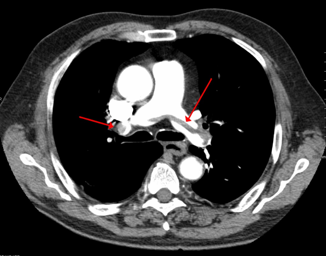
- Image with CT pulmonary angiography when the D-dimer is above the cutoff or probability is high.5
| PERC: all must be absent | |
|---|---|
| Age ≥ 50 | Hemoptysis |
| Heart rate ≥ 100 | Surgery or trauma needing general anesthesia within 4 weeks |
| Room-air SpO2 < 95% | Prior PE or DVT |
| Unilateral leg swelling | Hormone use (oral contraceptive, hormone replacement, estrogen) |
Apply PERC only when gestalt probability is low (under 15%).13
What the rule-out studies show
- PERC, 8,138 patients: low gestalt plus negative PERC applied to 20% of patients, with a 1.0% miss rate.3
- PROPER trial, 1,916 patients: a PERC strategy had 0.1% thromboembolic events at 3 months, cut CT use from 23% to 13%, and shortened ED stays by 36 minutes.4
- YEARS, 3,465 patients: three items (clinical signs of DVT, hemoptysis, PE the most likely diagnosis). The D-dimer cutoff is 1000 ng/mL with none of them and 500 ng/mL with any.14 Missed VTE at 3 months was 0.61%, and 14% fewer patients needed CT.5
- Age-adjusted D-dimer (ADJUST-PE, 3,346 patients): a cutoff of age × 10 µg/L over age 50.15 Patients between 500 and their age-adjusted cutoff had a 0.3% 3-month failure rate. In patients 75 and older, D-dimer excluded PE in 29.7% instead of 6.4%.6
- PEGeD, 2,017 patients: low clinical probability plus D-dimer under 1000 ng/mL had no VTE at follow-up, and chest imaging fell from 51.9% to 34.3%.7
- Pregnancy (pregnancy-adapted YEARS, 498 patients): no PE during follow-up, and CT avoided in 39% overall (65% in the first trimester, 32% in the third).8
Severity: AHA/ACC categories
| Category | Definition | Usual path |
|---|---|---|
| A | Asymptomatic, found incidentally (e.g. on cancer-staging CT) | Benefit of treatment still under study |
| B | Symptomatic, low severity score: PESI ≤ 85, sPESI 0, or Bova ≤ 4 | Many can go home on an oral anticoagulant |
| C | Symptomatic, higher score: PESI > 85, sPESI ≥ 1, or Bova > 4. C3 adds more RV imaging and biomarker abnormalities | Usually admitted; rarely benefit beyond anticoagulation |
| D | Incipient cardiopulmonary failure. D1 transient hypotension (e.g. syncope); D2 normotensive shock (e.g. raised lactate, AKI) | Consider advanced therapy (2b) |
| E | Persistent hypotension or cardiopulmonary failure. E1 cardiogenic shock; E2 refractory shock or arrest | Advanced therapy (2a) |
A respiratory modifier can be added to categories C–E. When respiratory and hemodynamic categories conflict, assign the higher one.2
sPESI (one point each)
| Item | Point if |
|---|---|
| Age | > 80 years |
| Cancer | history of |
| Chronic cardiopulmonary disease | history of |
| Heart rate | ≥ 110 |
| Systolic BP | < 100 mm Hg |
| SpO2 | < 90% |
Thresholds as listed by MDCalc.16 In the derivation study, sPESI 0 meant a 30-day mortality of 1.0% vs 10.9% for ≥ 1, and 1.1% vs 8.9% in a 7,106-patient validation cohort.9
Anticoagulation
- Hospitalized (categories C–E1): prefer LMWH over unfractionated heparin (Class 1), including for patients heading to catheter therapy or lysis.1,2
- Outpatients (A and B): prefer a DOAC over warfarin, including in obesity. Warfarin remains the choice in antiphospholipid syndrome and advanced liver disease.1,2
| Drug | PE treatment dose (FDA label) |
|---|---|
| Enoxaparin | 1 mg/kg SC every 12 h, or 1.5 mg/kg SC once daily (inpatient) |
| Apixaban | 10 mg PO twice daily × 7 days, then 5 mg twice daily |
| Rivaroxaban | 15 mg PO twice daily with food × 21 days, then 20 mg daily; avoid if CrCl < 15 |
Unreviewed doses. Verify against the current label and your institution's protocol before giving anything.
Doses from the FDA labels.11,12 Check renal function, weight and interactions; the apixaban label halves the dose with combined strong CYP3A4 and P-gp inhibitors.11
Advanced therapy
| Category | Systemic lysis, catheter-directed therapy, thrombectomy |
|---|---|
| E | Class 2a. The three are now treated as equivalent options; systemic lysis no longer comes first |
| D | Class 2b. May be considered; benefit unclear |
| C3 | Class 2b. Benefit unclear |
- Call the PE response team (PERT) for categories C–E (Class 1).1,2
- Alteplase dose (label): 100 mg IV over 2 hours. Start parenteral anticoagulation near the end of the infusion, once the PTT is no more than twice normal.17
- The bleeding trade-off (PEITHO, 1,005 intermediate-risk patients): tenecteplase cut death or decompensation (2.6% vs 5.6%) but raised major extracranial bleeding (6.3% vs 1.2%) and stroke (2.4% vs 0.2%). Day-30 mortality did not differ.10
Disposition and follow-up
- Category B: many patients can be discharged on an oral anticoagulant.1,2 sPESI 0 is a low-risk signal, but home care also needs a stable patient, safe anticoagulation and follow-up.16
- About 1 week after discharge: a visit or call to check access, bleeding and understanding (Class 1).1,2
- Within 3 months: follow-up for complications and persistent symptoms. If symptoms persist, test for chronic thromboembolic disease.1,2
- Duration: stop after 3–6 months for a first, strongly provoked PE without ongoing risk factors. Otherwise continued anticoagulation is generally preferred; reduced-dose apixaban or rivaroxaban can lower bleeding.2
Pitfalls
- Applying PERC to patients who aren't low risk by gestalt.13
- Ordering a D-dimer you will interpret with a fixed 500 cutoff in an 80-year-old.6
- Using a D-dimer to "rule out" PE in a high-probability patient. Go to imaging.5,7
- Reaching for lysis in category C. Most of these patients do not benefit beyond anticoagulation, and PEITHO showed the bleeding cost.2,10
- Discharging category B patients without a plan for anticoagulant access and early follow-up.1,2
How strong is the evidence?
The rule-out strategies are supported by large prospective studies and one randomized trial.3,4,5,6,7,8 Advanced therapy recommendations are mostly Class 2a–2b. The 2026 guideline is the first AHA/ACC acute PE guideline; its full text was not openly accessible, so recommendations here come from a published summary and are cited to both.1,2 For deeper reading, see LITFL and Emergency Medicine Cases.
On-shift teaching
Questions to ask a learner at the bedside. Tap a question to see the answer. Every answer uses facts cited above.
Q1Who can you apply PERC to?
Q2A low-probability 80-year-old has a D-dimer of 700 µg/L. Is PE ruled out?
Q3What are the YEARS items, and how do they change the D-dimer cutoff?
Q4What is category B, and what does it mean for disposition?
Q5Why not give lytics to every intermediate-risk PE?
Q6What is the apixaban dose for acute PE?
References
- 1Creager MA, Barnes GD, Giri J, et al. 2026 AHA/ACC/ACCP/ACEP/CHEST/SCAI/SHM/SIR/SVM/SVN Guideline for the Evaluation and Management of Acute Pulmonary Embolism in Adults. Circulation. 2026;153(12):e977–e1051.
- 2The 2026 AHA/ACC guideline for the management of acute pulmonary embolism. Endovascular Today, July 2026 supplement.
- 3Kline JA, Courtney DM, Kabrhel C, et al. Prospective multicenter evaluation of the pulmonary embolism rule-out criteria. J Thromb Haemost. 2008;6(5):772–780.
- 4Freund Y, Cachanado M, Aubry A, et al. Effect of the pulmonary embolism rule-out criteria on subsequent thromboembolic events among low-risk emergency department patients: the PROPER randomized clinical trial. JAMA. 2018;319(6):559–566.
- 5van der Hulle T, Cheung WY, Kooij S, et al. Simplified diagnostic management of suspected pulmonary embolism (the YEARS study). Lancet. 2017;390(10091):289–297.
- 6Righini M, et al. Age-adjusted D-dimer cutoff levels to rule out pulmonary embolism: the ADJUST-PE study. JAMA. 2014;311(11):1117–1124.
- 7Kearon C, de Wit K, Parpia S, et al. Diagnosis of pulmonary embolism with D-dimer adjusted to clinical probability (PEGeD). N Engl J Med. 2019;381(22):2125–2134.
- 8van der Pol LM, Tromeur C, Bistervels IM, et al. Pregnancy-adapted YEARS algorithm for diagnosis of suspected pulmonary embolism. N Engl J Med. 2019;380(12):1139–1149.
- 9Jiménez D, Aujesky D, Moores L, et al. Simplification of the pulmonary embolism severity index for prognostication in patients with acute symptomatic pulmonary embolism. Arch Intern Med. 2010;170(15):1383–1389.
- 10Meyer G, Vicaut E, Danays T, et al. Fibrinolysis for patients with intermediate-risk pulmonary embolism (PEITHO). N Engl J Med. 2014;370(15):1402–1411.
- 11Eliquis (apixaban) prescribing information. DailyMed, US National Library of Medicine.
- 12Xarelto (rivaroxaban) and Lovenox (enoxaparin) prescribing information. DailyMed, US National Library of Medicine.
- 13MDCalc. PERC Rule for Pulmonary Embolism.
- 14Cloyd T, Swaminathan A. YEARS. Core EM journal review; November 16, 2017.
- 15Wiki Journal Club. ADJUST-PE.
- 16MDCalc. Simplified PESI (Pulmonary Embolism Severity Index).
- 17Activase (alteplase) prescribing information. DailyMed, US National Library of Medicine.
AI-generated draft. Not reviewed by a physician. This page is for education only and may contain errors, including in doses. Do not use it to diagnose, treat or make decisions about any patient. Rely on your own clinical judgment, your institution's protocols and current drug labeling. Reading this page does not create a physician-patient relationship. Full terms