Wide-complex tachycardia
Bottom line
- Life-threatening features mean a synchronized shock now: shock, syncope with severe or ongoing hypotension, myocardial ischemia, severe heart failure, or just after ROSC.1,2
- Treat a regular wide-complex tachycardia as VT until proven otherwise. A history of myocardial infarction or heart failure predicted VT with over 95% positive predictive value, and no history feature reliably predicted SVT.3,4
- Never give verapamil to an undifferentiated wide-complex tachycardia. In one series, VT was misread as SVT in 39% of episodes, and every patient with misread VT who got verapamil deteriorated.5
- For stable monomorphic VT, procainamide beat amiodarone in the only randomized comparison: fewer major cardiac adverse events (9% vs 41%) and more terminations within 40 minutes (67% vs 38%).6
- Adenosine is a reasonable test only for a regular, monomorphic rhythm without pre-excitation. 90% of SVTs responded and 2% of VTs did, with no serious adverse events.1,7
First minutes
- Check for a pulse. No pulse: this is pulseless VT, so follow the cardiac arrest algorithm and defibrillate.1
- ABCDE. Monitor the ECG, blood pressure and SpO₂, record a 12-lead ECG, give oxygen if SpO₂ is below 94%, get IV access, and look for reversible causes.1
- Look for life-threatening features: shock (for example, systolic BP under 90 mm Hg with signs of poor perfusion), syncope, heart failure with pulmonary edema or raised JVP, or myocardial ischemia.1
- Unstable: synchronized cardioversion, up to 3 attempts, with sedation or anesthesia if the patient is conscious.1,2
- Stable: decide whether the QRS is regular and monomorphic, then follow the stable pathway below. Get expert help early.1
- No: stable
- No: regular
- No, or no response
- No
- Arrhythmia or features persist: get expert help. Look for ischemia and structural heart disease.1
VT or SVT with aberrancy?
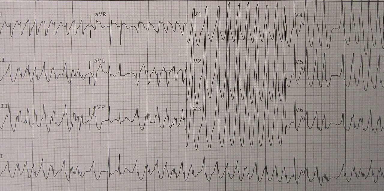
A regular wide-complex tachycardia can be VT, SVT with a bundle branch block, SVT slowed by a metabolic problem (hyperkalemia, sodium channel blockade, severe acidosis), antidromic AVRT in Wolff-Parkinson-White syndrome, or a paced rhythm.4 The ECG cannot always tell them apart.4
| Favors VT | Favors SVT with aberrancy |
|---|---|
| Prior MI, heart failure or recent angina (positive predictive value > 95%)3 | Old ECGs with the same bundle branch block morphology4 |
| Age over 35 (sensitivity 92%, positive predictive value 85%)3 | Old ECGs showing pre-excitation4 |
| AV dissociation, capture beats or fusion beats4 | Past episodes stopped by adenosine or vagal maneuvers4 |
| Precordial concordance (all positive or all negative)4 | A typical RBBB or LBBB pattern4 |
| QRS > 160 ms, or a northwest axis4 | — |
No history feature strongly predicted SVT. Age 35 or under was the best, with a positive predictive value of only 70%.3
ECG algorithms
- Brugada criteria, in order: no RS complex in any precordial lead; an RS interval over 100 ms; AV dissociation; then VT morphology in V1–V2 and V6. Any yes means VT.4
- Lead aVR (Vereckei): a dominant initial R wave in aVR points to VT.4
- None is good enough to rule VT out. In 3,966 patients across 14 studies, the algorithms pooled to 89% sensitivity and 71% specificity for VT.11
- In a head-to-head comparison of 260 tracings, all five methods had only moderate accuracy. Newer methods were no better than the Brugada algorithm (accuracy 77.5%).12
In 46 emergency episodes, every SVT with aberrancy was diagnosed correctly, but 15 of 38 VTs (39%) were called SVT. AV dissociation was visible in 73% of the misread ECGs. Verapamil was given in 13 of those 15 episodes, and all 13 deteriorated.5
Treatment
| Treatment | Adult dose | Use for | Notes |
|---|---|---|---|
| Electrical | |||
| Synchronized cardioversion | VT: 120–150 J first, then step up | Unstable patients. Also the UK first choice for stable monomorphic VT with structural or unknown heart disease | Sedate or anesthetize if conscious, and expect sedation to worsen hemodynamics.1 |
| Regular, monomorphic: drugs | |||
| Procainamide | 10–15 mg/kg IV over 20 min (max 1 g) | Stable VT; first-line drug in the UK algorithm | Label: no faster than 50 mg/min; stop for hypotension, persistent conduction problems, or QRS widening of 50%. Contraindicated in torsades and lupus.1,8 |
| Amiodarone | 300 mg IV over 10–60 min, then 900 mg over 24 h | Stable VT if procainamide is unavailable or contraindicated | US label load: 150 mg over 10 min, then 1 mg/min for 6 h, then 0.5 mg/min. Hypotension in 16% in label trials.1,13 |
| Adenosine | 6 mg rapid IV, then 12 mg, then 18 mg | Regular, monomorphic rhythm that may be SVT, with no pre-excitation | US label: 6 mg, then 12 mg, repeated once. Avoid in bronchospasm.1,14 |
| Polymorphic VT with a long QT (torsades) | |||
| Magnesium | 8 mmol IV over 10 min | Torsades | Avoid amiodarone. Consider isoprenaline or pacing to raise the heart rate.1 |
| Avoid | |||
| Verapamil or diltiazem | Do not give | Undifferentiated wide-complex tachycardia | Hemodynamic deterioration in every misdiagnosed VT treated with verapamil.5 |
| Lidocaine | Not first-line | — | Terminated 3 of 14 VTs vs 12 of 15 with procainamide.15 |
Unreviewed doses. Verify against the current label and your institution's protocol before giving anything.
For a stable patient in monomorphic VT, the UK guideline recommends electrical cardioversion when there is structural heart disease or the heart's state is unknown, and keeps drugs for patients in whom sedation is too risky.1,2 The 2025 American Heart Association guideline, as summarized, suggests vagal maneuvers and drug therapy for stable wide-complex tachycardia.9,10 Either way, shock if the patient becomes unstable.1
Procainamide or amiodarone?
- PROCAMIO (n = 62 analyzed, tolerated wide-complex tachycardia, probably VT): procainamide 10 mg/kg over 20 min vs amiodarone 5 mg/kg over 20 min. Major cardiac adverse events within 40 min: 9% vs 41%. Termination within 40 min: 67% vs 38%. In structural heart disease, adverse events were 11% vs 43%.6
- Amiodarone terminates VT poorly. In an ED series it stopped 8 of 28 VTs (29%) within 20 minutes.16 In another, it stopped 15% within 20 minutes and 29% within an hour; 17% needed emergency cardioversion for hemodynamic deterioration.17
- A systematic review (5 small studies) found procainamide, ajmaline and sotalol all better than lidocaine. Amiodarone was not more effective than procainamide.18
- Lidocaine: in a randomized trial, procainamide stopped 12 of 15 VTs and lidocaine 3 of 14.15
Adenosine
- In 197 ED patients with a regular wide-complex tachycardia, 90% of SVTs and 2% of VTs responded to adenosine. A response raised the odds of SVT 36-fold. No patient needed emergency treatment for an adenosine side effect.7
- Use it only for a regular, monomorphic rhythm. The UK algorithm gives it only when there is no pre-excitation, and says to treat as VT if it fails.1
- Adenosine does not convert VT. Rare ventricular fibrillation has been reported, mostly with digoxin.14
Torsades
In 12 patients with torsades, a 2 g magnesium sulfate bolus abolished it in 9, and a second bolus worked in the other 3. Magnesium did not help polymorphic VT with a normal QT. Most episodes were caused by antiarrhythmic drugs, and 8 patients had potassium below 3.5 mEq/L.19
After the rhythm is controlled
- Persistent arrhythmias often mean structural heart disease or unresolved problems such as myocardial ischemia. Look for both.1
- Get expert help if the arrhythmia or life-threatening features persist.1
- Watch the blood pressure after amiodarone. Hypotension is its most common adverse reaction, usually in the first hours and related to the infusion rate. Treat it first by slowing the infusion.13
- Watch for bradycardia and AV block after amiodarone; some patients need pacing.13
Pitfalls
- Calling it SVT without looking for AV dissociation. It was visible on 73% of the ECGs of misread VT.5
- Giving verapamil or diltiazem before VT is excluded.5
- Giving adenosine for an irregular wide-complex rhythm, or with pre-excitation.1
- Trusting one ECG algorithm to rule VT out. Specificity is only moderate.11,12
- Treating torsades with amiodarone or procainamide. Use magnesium.1,8
- Missing a metabolic cause, such as hyperkalemia or sodium channel blocker toxicity, that widens the QRS of an SVT.4
How strong is the evidence?
Most of the evidence is small and old. Only one randomized trial compares the two main drugs, and it analyzed 62 patients.6 The systematic review of drug therapy found quality problems in all five studies.18 Recommendations on cardioversion versus drugs for stable VT differ between the UK and US guidelines.1,9 Neither the full ERC nor the full AHA guideline text could be read for this page.2,9
On-shift teaching
Questions to ask a learner at the bedside. Tap a question to see the answer. Every answer uses facts cited above.
Q1Why is verapamil dangerous in a wide-complex tachycardia?
Q2Which parts of the history make VT likely?
Q3Can an ECG algorithm rule VT out?
Q4Is adenosine safe in a regular wide-complex tachycardia?
Q5Procainamide or amiodarone for stable VT?
References
What this page is built on
19 sources, cited 96 times. Dots are sources, filled by how far each was read; stronger color means a larger share of the citations. Tap a tier to see its sources.
Checked by the writer, not a physician. The dots show how each source was read, not that anyone has confirmed the claims.
- 1Resuscitation Council UK. Adult advanced life support guidelines (2025 Resuscitation Guidelines, adapted from the ERC 2025 guidelines), published October 27, 2025; and Adult tachyarrhythmia algorithm, updated March 2026.
- 2Soar J, Böttiger BW, Carli P, et al. European Resuscitation Council Guidelines 2025 Adult Advanced Life Support. Resuscitation. 2025;215 Suppl 1:110769.
- 3Baerman JM, Morady F, DiCarlo LA Jr, de Buitleir M. Differentiation of ventricular tachycardia from supraventricular tachycardia with aberration: value of the clinical history. Ann Emerg Med. 1987;16(1):40–43.
- 4Burns E, Buttner R. VT versus SVT. Life in the Fast Lane ECG library; updated September 24, 2026.
- 5Stewart RB, Bardy GH, Greene HL. Wide complex tachycardia: misdiagnosis and outcome after emergent therapy. Ann Intern Med. 1986;104(6):766–771.
- 6Ortiz M, Martín A, Arribas F, et al.; PROCAMIO Study Investigators. Randomized comparison of intravenous procainamide vs. intravenous amiodarone for the acute treatment of tolerated wide QRS tachycardia: the PROCAMIO study. Eur Heart J. 2017;38(17):1329–1335.
- 7Marill KA, Wolfram S, Desouza IS, et al. Adenosine for wide-complex tachycardia: efficacy and safety. Crit Care Med. 2009;37(9):2512–2518.
- 8Procainamide hydrochloride injection. FDA prescribing information (RemedyRepack). DailyMed.
- 9Wigginton JG, Agarwal S, Bartos JA, et al. Part 9: Adult Advanced Life Support: 2025 American Heart Association Guidelines for Cardiopulmonary Resuscitation and Emergency Cardiovascular Care. Circulation. 2025;152(16 Suppl 2):S538–S577.
- 10McKinnon M. 2025 AHA Guidelines Update: Key Changes in CPR and ECC. EM Ottawa Blog; January 8, 2026.
- 11Sun X, Teng Y, Mu S, Wang Y, Chen H. Diagnostic accuracy of different ECG-based algorithms in wide QRS complex tachycardia: a systematic review and meta-analysis. BMJ Open. 2023;13(7):e069273.
- 12Jastrzebski M, Kukla P, Czarnecka D, Kawecka-Jaszcz K. Comparison of five electrocardiographic methods for differentiation of wide QRS-complex tachycardias. Europace. 2012;14(8):1165–1171.
- 13Amiodarone hydrochloride injection. FDA prescribing information (Henry Schein). DailyMed.
- 14Adenosine injection. FDA prescribing information (Henry Schein). DailyMed.
- 15Gorgels AP, van den Dool A, Hofs A, et al. Comparison of procainamide and lidocaine in terminating sustained monomorphic ventricular tachycardia. Am J Cardiol. 1996;78(1):43–46.
- 16Marill KA, deSouza IS, Nishijima DK, et al. Amiodarone is poorly effective for the acute termination of ventricular tachycardia. Ann Emerg Med. 2006;47(3):217–224.
- 17Tomlinson DR, Cherian P, Betts TR, Bashir Y. Intravenous amiodarone for the pharmacological termination of haemodynamically-tolerated sustained ventricular tachycardia: is bolus dose amiodarone an appropriate first-line treatment? Emerg Med J. 2008;25(1):15–18.
- 18deSouza IS, Martindale JL, Sinert R. Antidysrhythmic drug therapy for the termination of stable, monomorphic ventricular tachycardia: a systematic review. Emerg Med J. 2015;32(2):161–167.
- 19Tzivoni D, Banai S, Schuger C, et al. Treatment of torsade de pointes with magnesium sulfate. Circulation. 1988;77(2):392–397.
AI-generated draft. Not reviewed by a physician. This page is for education only and may contain errors, including in doses. Do not use it to diagnose, treat or make decisions about any patient. Rely on your own clinical judgment, your institution's protocols and current drug labeling. Reading this page does not create a physician-patient relationship. Full terms