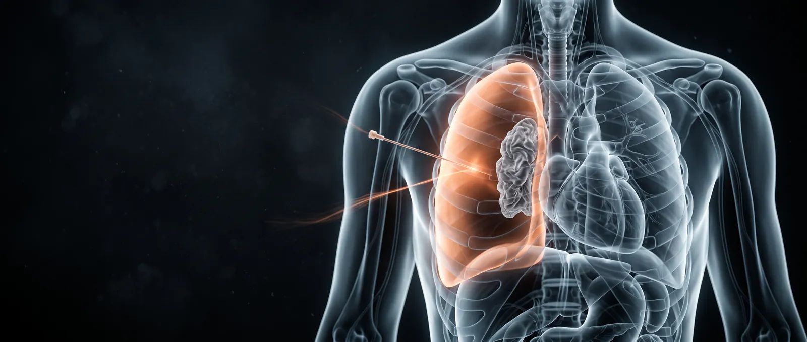
Tension pneumothorax
Bottom line
- Decompress before imaging only for hemodynamic instability or severe respiratory compromise. Otherwise, confirm it first.1
- Ventilated patients crash fast. Compared with patients breathing on their own, they had 12.6 times the odds of hypotension and 17.7 times the odds of cardiac arrest, usually within minutes.2
- Open thoracostomy over the needle if you have the skills, followed by a chest drain.1 On imaging, needles failed to reach the pleural space in about a third of cases.3
- If you use a needle, use a long one. Each extra centimeter of length lowered the failure rate by about 8 percentage points. A 2025 meta-analysis suggests 7 cm.3
- Bedside ultrasound beats the supine chest film: sensitivity 91% vs 47% for traumatic pneumothorax.4 A negative eFAST still does not exclude it.1
Recognize
- Breathing on their own: hypoxia was reported in 50% of cases, and 9.3% progressed to respiratory arrest.2
- On a ventilator: hypoxia was reported in 91.8%. Odds of hypotension were 12.6 times higher and odds of cardiac arrest 17.7 times higher. In one cohort, 39.6% of ventilated patients had no pulse at diagnosis, against none of those breathing on their own. Of those who became hypotensive or arrested, 70.4% did so within minutes.2
- Think of it in a ventilated patient who suddenly deteriorates, and after central line placement.5
- Classic signs are hard to elicit in a busy resuscitation: reduced breath sounds, a hyper-resonant chest and tracheal deviation.3
- A wrong diagnosis has a cost. A needle in a chest without tension can cause a pneumothorax.3 In one prehospital series, at least 39% of decompressed patients had no tension pneumothorax on CT.6
First minutes
- Unstable or in severe respiratory compromise: decompress now, before imaging.1
- Use open thoracostomy if you have the skills, rather than a needle. In a patient breathing on their own, place the chest drain through the thoracostomy.1
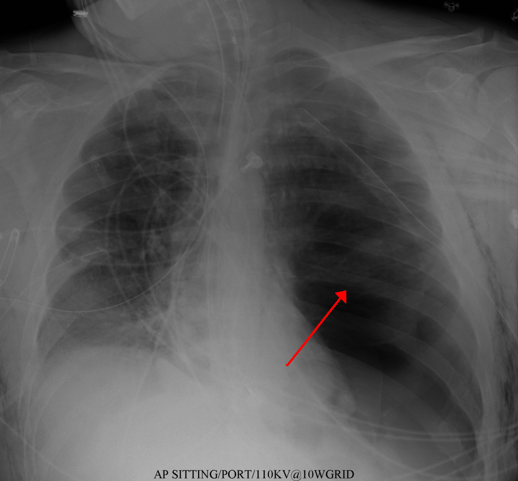
- Stable: confirm first. Use chest X-ray or eFAST in the primary survey for severe respiratory compromise. Consider CT for patients who are stable or responding to resuscitation.1
- Watch for recurrence after decompression.1
- Open chest wound: cover it with a simple occlusive dressing and watch for tension.1
- On a ventilator?YesExpect a fast crash. Hypotension or arrest often comes within minutes.2
- Either wayHemodynamic instability or severe respiratory compromise?NoImage first: chest X-ray or eFAST, or CT if stable. A negative eFAST does not exclude pneumothorax.1
- YesDecompress now, before imaging
- Chest drain next, through the thoracostomy in a patient breathing on their own.1
Decompression
| ATLS | European Trauma Course | 2025 meta-analysis | |
|---|---|---|---|
| Site | 4th or 5th intercostal space, anterior midaxillary line | 2nd intercostal space, midclavicular line | Right: 2nd space midclavicular or 5th space midaxillary. Left: 2nd space midclavicular only |
| Needle | 5 cm for smaller adults, 8 cm for larger adults | 14 or 16 gauge "extra-long" cannula | 7 cm |
Course recommendations as summarized in the 2025 meta-analysis, which made the suggestions in the last column.3
| Site (5 cm needle) | Chest wall thickness | Failure rate |
|---|---|---|
| 2nd intercostal space, midclavicular line | 42.8 mm | 38% |
| 4th/5th space, midaxillary line | 39.9 mm | 31% |
| 4th/5th space, anterior axillary line | 34.3 mm | 13% |
Pooled from observational studies in a 2016 meta-analysis.7
Where to put the needle. The 2016 meta-analysis found the lowest predicted failure at the 4th/5th space in the anterior axillary line.7 The 2025 meta-analysis found no significant difference between sites once needle length was matched (p = 0.052). On the left, the risk of heart injury at the 5th space rose with needle length, so it suggests the 2nd space in the midclavicular line on that side.3 In a CT review, an 8 cm catheter at the 5th space in the anterior axillary line put the heart at risk in 42% of patients.6
- Needles fail often. On imaging, 32.8% failed to enter the pleural space (24 studies, 8,046 patients). Clinical improvement failed in about half.3
- In one prehospital CT review, 39% to 76% of needles at the 2nd space failed to reach the pleura, depending on the method. Injured chest walls were thicker than uninjured ones.6
- Finger or needle? Studies of paramedic decompression were mostly retrospective, with no randomized trials. Complications were reported in 13.7% after needle and 4.8% after finger thoracostomy. Mortality ranges were too wide to compare.8
- NICE prefers open thoracostomy when the expertise is there.1 REBEL EM argues finger thoracostomy is faster and more reliable than the needle.5
Imaging
Pitfalls
- Waiting for a chest film in an unstable patient.1
- Trusting a short needle at the 2nd intercostal space in a thick chest wall.7,3
- Placing a long needle low on the left, near the heart.3,6
- Assuming a needle that "worked" has fixed the problem. Place a drain and watch for recurrence.1
- Calling it tension when it isn't. At least 39% of one prehospital series had no tension pneumothorax.6
- Forgetting it in a ventilated patient who suddenly becomes hypotensive.2,5
How strong is the evidence?
Weak and observational. In the 2025 meta-analysis, 50 of 51 studies were level IV evidence, and heterogeneity was extreme (I² > 99%).3 The review of paramedic decompression found no randomized trials.8 What we know about the clinical picture comes from 5 cohort studies and 156 case reports and series.2 The NICE recommendations were published in 2016.1
On-shift teaching
Questions to ask a learner at the bedside. Tap a question to see the answer. Every answer uses facts cited above.
Q1Why is tension pneumothorax more dangerous in a ventilated patient?
Q2When do you decompress before imaging?
Q3Why do needles at the 2nd intercostal space fail?
Q4Left-sided tension and only a long needle: where do you put it?
Q5How does bedside ultrasound compare with a supine chest X-ray for traumatic pneumothorax?
Q6You've done a finger thoracostomy. What next?
References
What this page is built on
9 sources, cited 62 times. Dots are sources, filled by how far each was read; stronger color means a larger share of the citations. Tap a tier to see its sources.
Checked by the writer, not a physician. The dots show how each source was read, not that anyone has confirmed the claims.
- 1National Institute for Health and Care Excellence. Major trauma: assessment and initial management (NG39). February 2016.
- 2Roberts DJ, Leigh-Smith S, Faris PD, et al. Clinical presentation of patients with tension pneumothorax: a systematic review. Ann Surg. 2015;261(6):1068–1078.
- 3Ahmad SJS, Degiannis JR, Head M, et al. Meta-analysis of the optimal needle length and decompression site for tension pneumothorax and consensus recommendations on current ATLS and ETC guidelines. World J Emerg Surg. 2025;20(1):39.
- 4Chan KK, Joo DA, McRae AD, et al. Chest ultrasonography versus supine chest radiography for diagnosis of pneumothorax in trauma patients in the emergency department. Cochrane Database Syst Rev. 2020;7(7):CD013031.
- 5Swaminathan A. REBEL Core Cast 139.0: Pneumothorax decompression. REBEL EM; August 18, 2025.
- 6Lesperance RN, Carroll CM, Aden JK, Young JB, Nunez TC. Failure rate of prehospital needle decompression for tension pneumothorax in trauma patients. Am Surg. 2018;84(11):1750–1755.
- 7Laan DV, Vu TD, Thiels CA, et al. Chest wall thickness and decompression failure: a systematic review and meta-analysis comparing anatomic locations in needle thoracostomy. Injury. 2016;47(4):797–804.
- 8Sharrock MK, Shannon B, Garcia Gonzalez C, et al. Prehospital paramedic pleural decompression: a systematic review. Injury. 2021;52(10):2778–2786.
- 9Alrajab S, Youssef AM, Akkus NI, Caldito G. Pleural ultrasonography versus chest radiography for the diagnosis of pneumothorax: review of the literature and meta-analysis. Crit Care. 2013;17(5):R208.
AI-generated draft. Not reviewed by a physician. This page is for education only and may contain errors, including in doses. Do not use it to diagnose, treat or make decisions about any patient. Rely on your own clinical judgment, your institution's protocols and current drug labeling. Reading this page does not create a physician-patient relationship. Full terms