
Necrotizing soft tissue infection
Bottom line
- Suspect it early. Most cases are first diagnosed as cellulitis. Swelling, redness and pain out of proportion to the exam should raise the alarm.1
- Call the surgeon on suspicion, not proof. Surgery within 6 hours of arrival was linked to 19% mortality, against 32% when it came later (OR 0.43).2
- No single finding rules it out. Fever was present in only 46% and hypotension in 21%. A LRINEC score ≥ 6 missed about a third of cases (sensitivity 68%).3
- Imaging must not delay the operating room. CT is the best ED test (sensitivity 89%, specificity 93%). Plain films miss about half of cases.3,1
- Antibiotics: broad, plus a toxin blocker. Cover MRSA, Gram-negatives and anaerobes, and include clindamycin or linezolid.1,4
Recognize
- Common local signs: swelling (81% of cases), pain or tenderness (79%), erythema (71%), warmth (44%), bullae (26%), skin necrosis (24%) and crepitus (20%).1
- Late signs: hemorrhagic bullae, crepitus, skin necrosis, dusky discoloration, wood-hard tissue and numb skin. Deep tissue is involved first, so the skin can look deceptively mild.5
- Red flags: pain or swelling beyond the edge of the redness, systemic illness, and rapid spread.5 Treat a rapidly progressing soft tissue infection as necrotizing from the start. It can worsen within hours.1
- Risk factors: diabetes (up to 44.5% of patients), kidney disease, arterial disease, injection drug use, BMI > 30, liver disease, immunosuppression, recent surgery, and skin breaks as minor as insect bites or injection sites.1
- Ask about water. Seawater suggests Vibrio vulnificus, especially with liver disease. Fresh or brackish water, soil or wood suggests Aeromonas.1
| Type | Organisms | Typical setting |
|---|---|---|
| I · polymicrobial | Aerobes and anaerobes | Bowel surgery, perineal or perianal abscess, pressure ulcers, injection sites1 |
| II · monomicrobial | Group A strep, S. aureus; Vibrio, Aeromonas | Community-acquired, mostly limbs; about two-thirds in the legs1 |
| III · gas gangrene | Clostridium (mostly C. perfringens) | Trauma, or spontaneous; spreads within hours1 |
Fournier gangrene is an NSTI of the genitals or perineum, usually from an anorectal, urinary or skin source.1 The approach to diagnosis, antibiotics and surgery is similar for every type.1
First hour
- Call the surgeon now for suspected NSTI, systemic toxicity or suspected gas gangrene.4,1
- Resuscitate as for sepsis. Give 1 L of balanced crystalloid over 30 minutes, and up to 30 mL/kg within 3 hours if hypotensive or lactate is high. Start norepinephrine for a MAP under 65, peripherally if needed.1
- Give antibiotics within 1 hour. Use a broad regimen that includes clindamycin or linezolid (table below).1,4
- Draw blood cultures. Deep tissue samples taken during the first debridement identify the cause in most cases.1
- Image only if it won't delay surgery.1
- Resuscitate: 1 L balanced crystalloid over 30 min, up to 30 mL/kg within 3 h if hypotensive or lactate is high, norepinephrine for MAP < 65.1
- Uncertain
- NoAdmit and re-examine often. The picture can change within hours. Call the surgeon back if it spreads.1
- After surgery: ICU care, a re-look every 12–24 h, and antibiotics narrowed to the cultures.1
Tests and scores
| Finding or test | Sensitivity | Specificity |
|---|---|---|
| Examination | ||
| Fever | 46.0% | 77.0% |
| Hemorrhagic bullae | 25.2% | 95.8% |
| Hypotension | 21.0% | 97.7% |
| Imaging | ||
| Plain radiograph | 48.9% | 94.0% |
| CT | 88.5% | 93.3% |
| Score | ||
| LRINEC ≥ 6 | 68.2% | 84.8% |
| LRINEC ≥ 8 | 40.8% | 94.9% |
Pooled from 23 studies (n = 5,982) in adults with a soft tissue infection worrying for NSTI, confirmed at surgery or by histology.3
LRINEC score
| Lab | 0 points | Points |
|---|---|---|
| C-reactive protein | < 150 mg/L | ≥ 150: 4 |
| White cells (×1,000/µL) | < 15 | 15–25: 1 · > 25: 2 |
| Hemoglobin (g/dL) | > 13.5 | 11–13.5: 1 · < 11: 2 |
| Sodium (mEq/L) | ≥ 135 | < 135: 2 |
| Creatinine (mg/dL) | ≤ 1.6 | > 1.6: 2 |
| Glucose (mg/dL) | ≤ 180 | > 180: 1 |
Items from MDCalc.6 The derivation study used a cutoff of 6.9
In its retrospective derivation, a score of 6 or more had a negative predictive value of 96%.9 Pooled across later studies, LRINEC ≥ 6 had a sensitivity of only 68%.3 The WSES pathways call its accuracy poor, and say a low score does not rule out NSTI.1 MDCalc advises skipping the score when clinical suspicion is high and going straight to surgery.6
Imaging
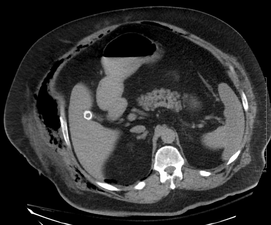
- CT: look for fat stranding, fluid and gas tracking along fascial planes, and fascia that is thickened or does not enhance with contrast.1
- Plain X-ray should not be used to rule out NSTI.1
- MRI is hard to do in an emergency and is not the first-choice test.1
- Bedside ultrasound: in a review of 3 studies (n = 221), sensitivity ranged from 85.4% to 100% and specificity from 44.7% to 98.2%. Fluid along the fascia was the most sensitive sign (85.4%), and subcutaneous gas the most specific (100%).7
- When suspicion is high, imaging must not delay or deter surgery.1
Antibiotics
| Agent | Adult dose (normal renal function) | Notes |
|---|---|---|
| Gram-negative and anaerobic cover (one of) | ||
| Piperacillin-tazobactam | 4.5 g IV every 6 h | WSES dose for unstable patients. IDSA lists 3.37 g every 6–8 h.1,4 |
| Meropenem | 1 g IV every 8 h | Preferred where ESBL-producing organisms are common.1,4 |
| Ceftriaxone + metronidazole | 2 g IV daily + 500 mg IV every 8 h | WSES option for stable patients.1 |
| MRSA cover (one of) | ||
| Vancomycin | 30 mg/kg/day IV in 2 divided doses | WSES gives a 25–30 mg/kg load. Adjust to levels and kidney function.4,1 |
| Linezolid | 600 mg IV every 12 h | Also blocks toxin production.1,4 |
| Toxin suppression | ||
| Clindamycin | 600–900 mg IV every 8 h | Add it unless linezolid is the MRSA drug.1,4 |
| Directed therapy | ||
| Group A strep: penicillin + clindamycin | 2–4 million units IV every 4–6 h + 600–900 mg IV every 8 h | For documented streptococcal NSTI.4 |
| Vibrio: doxycycline + ceftriaxone or cefotaxime | Doxycycline 100 mg IV every 12 h | Seawater exposure.4,1 |
| Aeromonas: doxycycline + ciprofloxacin or ceftriaxone | Doxycycline 100 mg IV every 12 h | Fresh water or soil exposure.4,1 |
Unreviewed doses. Verify against the current label and your institution's protocol before giving anything.
Continue antibiotics until no further debridement is needed, then narrow them to the culture results.1 Set doses by the patient and the drug, with pharmacy input.1
- Why clindamycin or linezolid: both can markedly reduce exotoxin production by Gram-positive organisms.1 In 1,956 US inpatients with invasive streptococcal infection on a beta-lactam, adding clindamycin in group A infection was linked to lower in-hospital mortality (6.5% vs 11.0%, adjusted OR 0.44).10
- IVIG: in the INSTINCT trial, 100 ICU patients with NSTI received 25 g of IVIG or placebo daily for 3 days. Physical function at 6 months did not differ.11
- Hyperbaric oxygen: there are no randomized trials. Use it only if available on site, and never transfer for it or let it delay surgery or ICU care.1
Surgery and disposition
- Time matters. Across 109 studies (6,051 patients), 21.1% died. Surgery within 6 hours of arrival was linked to lower mortality (OR 0.43; 19% vs 32%), and so was surgery within 12 hours (OR 0.41).2
- Delays before arrival did not change mortality, and no timing measure reduced amputations.2
- Debride in the operating room and remove all necrotic tissue, which can mean amputation.1,12
- Re-look every 12–24 hours, or sooner if the patient worsens, until little or no further debridement is needed.1
- ICU: about half of patients develop septic shock.1 Care needs surgeons, intensivists and infectious disease specialists working together.12
- Fournier gangrene: a Fournier's Gangrene Severity Index above 9 predicts death.1
Pitfalls
- Calling it cellulitis and sending the patient home. Most NSTIs are first diagnosed that way.1
- Using a low LRINEC score, no fever or a normal X-ray to rule it out.3,6
- Waiting for CT or MRI when suspicion is already high.1
- Leaving out the toxin blocker (clindamycin or linezolid).1,4
- Missing the water history that points to Vibrio or Aeromonas.1
- Being reassured by mild-looking skin. Deep tissue is involved first.5
- Transferring a patient for hyperbaric oxygen.1
How strong is the evidence?
The IDSA rates its NSTI recommendations strong but based on low-quality evidence.4 The 6-hour surgery target comes from a meta-analysis of 33 studies with 2,123 patients, not from trials.2 LRINEC was derived retrospectively.9 Support for clindamycin is from a retrospective cohort.10 The only randomized trial of IVIG was negative.11 NSTIs are commonly missed.8
On-shift teaching
Questions to ask a learner at the bedside. Tap a question to see the answer. Every answer uses facts cited above.
Q1How sensitive are fever and hypotension for NSTI?
Q2The LRINEC score is 4, but the patient has pain far beyond the redness. What next?
Q3How much does the time to surgery matter?
Q4Why add clindamycin or linezolid to a broad regimen?
Q5A patient with cirrhosis has a fast-spreading leg infection after cleaning fish at the beach. What organism, and what antibiotics?
References
What this page is built on
12 sources, cited 106 times. Dots are sources, filled by how far each was read; stronger color means a larger share of the citations. Tap a tier to see its sources.
Checked by the writer, not a physician. The dots show how each source was read, not that anyone has confirmed the claims.
- 1Sartelli M, Coccolini F, Kluger Y, et al. WSES/GAIS/WSIS/SIS-E/AAST global clinical pathways for patients with skin and soft tissue infections. World J Emerg Surg. 2022;17(1):3.
- 2Nawijn F, Smeeing DPJ, Houwert RM, Leenen LPH, Hietbrink F. Time is of the essence when treating necrotizing soft tissue infections: a systematic review and meta-analysis. World J Emerg Surg. 2020;15:4.
- 3Fernando SM, Tran A, Cheng W, et al. Necrotizing soft tissue infection: diagnostic accuracy of physical examination, imaging, and LRINEC score: a systematic review and meta-analysis. Ann Surg. 2019;269(1):58–65.
- 4Stevens DL, Bisno AL, Chambers HF, et al. Practice guidelines for the diagnosis and management of skin and soft tissue infections: 2014 update by the Infectious Diseases Society of America. Clin Infect Dis. 2014;59(2):e10–e52.
- 5Berger B, Hill J. Necrotizing fasciitis and the spectrum of soft tissue infections. Taming the SRU; September 3, 2018.
- 6MDCalc. LRINEC Score for Necrotizing Soft Tissue Infection.
- 7Marks A, Patel D, Sundaram T, Johnson J, Gottlieb M. Ultrasound for the diagnosis of necrotizing fasciitis: a systematic review of the literature. Am J Emerg Med. 2023;65:31–35.
- 8Pelletier J, Gottlieb M, Long B, et al. Necrotizing soft tissue infections (NSTI): pearls and pitfalls for the emergency clinician. J Emerg Med. 2022;62(4):480–491.
- 9Wong CH, Khin LW, Heng KS, Tan KC, Low CO. The LRINEC (Laboratory Risk Indicator for Necrotizing Fasciitis) score: a tool for distinguishing necrotizing fasciitis from other soft tissue infections. Crit Care Med. 2004;32(7):1535–1541.
- 10Babiker A, Li X, Lai YL, et al. Effectiveness of adjunctive clindamycin in β-lactam antibiotic-treated patients with invasive β-haemolytic streptococcal infections in US hospitals: a retrospective multicentre cohort study. Lancet Infect Dis. 2021;21(5):697–710.
- 11Madsen MB, Hjortrup PB, Hansen MB, et al. Immunoglobulin G for patients with necrotising soft tissue infection (INSTINCT): a randomised, blinded, placebo-controlled trial. Intensive Care Med. 2017;43(11):1585–1593.
- 12Sartelli M, Coccolini F, Labricciosa FM, et al. Necrotizing soft-tissue infections survival guide in adult patients: a position statement by the Global Alliance for Infections in Surgery. J Trauma Acute Care Surg. 2026;100(2):320–331.
AI-generated draft. Not reviewed by a physician. This page is for education only and may contain errors, including in doses. Do not use it to diagnose, treat or make decisions about any patient. Rely on your own clinical judgment, your institution's protocols and current drug labeling. Reading this page does not create a physician-patient relationship. Full terms