
Hyperkalemia
Bottom line
- Read the ECG, not just the number. In severe hyperkalemia, QRS widening, heart rate under 50, and junctional rhythm predicted short-term adverse events. Peaked T waves did not.3 A normal ECG does not rule out danger.4
- ECG changes mean IV calcium first. It works in minutes, wears off within about an hour, and does not lower potassium.1
- Shift potassium with insulin plus glucose and nebulized albuterol 10–20 mg. Expect a drop of roughly 0.7–1.2 mEq/L.1,5
- Hypoglycemia is the most common treatment harm. It typically appears around 2 hours after insulin, so check glucose for 6 hours.1,12
- Remove potassium with a binder (SZC) or dialysis. Sodium polystyrene sulfonate (Kayexalate) is slow, unpredictable, and linked to bowel injury.1,16
Hyperkalemia in 30 seconds
Every spoken line and on-screen number comes from a cited line in this article. The pictures are AI illustrations, not clinical images.
Severity
Bands from the UK Kidney Association, which uses the European Resuscitation Council definition.1 No single treatment threshold is agreed in US emergency practice.2 For potassium, 1 mmol/L equals 1 mEq/L.
First 10 minutes
- Monitor and get a 12-lead ECG. Get an ECG for any potassium ≥ 6.0. Use continuous cardiac monitoring for ≥ 6.5, for any ECG changes, or for 6.0–6.4 in a patient who is unwell or whose level is rising.1
- Confirm fast. Run potassium on a blood gas analyzer while the lab result is pending.1 A high level with a normal ECG should raise the question of a spurious result.19
- ECG changes or instability: give IV calcium now.1,19
- Shift: insulin with glucose, plus nebulized albuterol for severe hyperkalemia.1
- Plan removal and find the cause. Start a binder. In anuric or dialysis patients, call for dialysis early.19,1
- Recheck potassium at 1, 2, 4, 6 and 24 hours, aiming for < 6.0 within 2 hours. Follow the glucose schedule below.1
- Then, for everyoneShift: regular insulin 10 units IV with 25 g glucose, plus nebulized albuterol 10–20 mg.1
- Pre-treatment glucose under 126 mg/dL?YesRun D10 at 50 mL/h for 5 hours.1
- Either wayCheck glucose at 0, 30, 60, 90, 120, 180, 240, 300 and 360 minutes after insulin.1
- NoRemove: SZC 10 g PO three times daily for K ≥ 6.5; consider it for 6.0–6.4.1
- Recheck potassium at 1, 2, 4, 6 and 24 hours, aiming for < 6.0 within 2 hours.1
ECG
Typical changes are peaked T waves, flattened P waves with a longer PR interval, a widening QRS, and finally a sine wave and arrest. They do not always appear in that order, and several can appear at once.1 See the LITFL ECG library for more examples.17
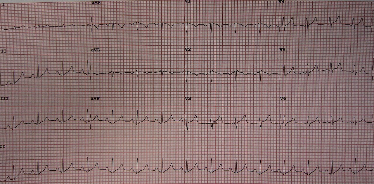
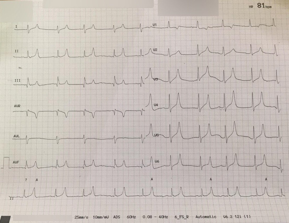
| Finding (K ≥ 6.5, n = 188) | Adverse events within 6 h |
|---|---|
| Heart rate < 50 | RR 12.3 (6.7–22.6) |
| Junctional rhythm | RR 7.5 (5.3–11.1) |
| QRS prolongation | RR 4.7 (2.0–11.2) |
| Peaked T waves | RR 0.77 (0.35–1.70), not significant |
Single-center retrospective study. Adverse events (mostly symptomatic bradycardia) occurred in 15% of patients. Every patient who had one also had at least one hyperkalemic finding on the preceding ECG.3
The ECG is insensitive. In 90 admitted patients with potassium ≥ 6, the ECG missed many cases, and readers agreed only moderately. The authors advise managing by the clinical picture and serial potassium levels, not by the ECG alone.4
Treatment
| Agent | Adult dose | Onset | Lasts | Notes |
|---|---|---|---|---|
| 1 · Protect the heart | ||||
| Calcium gluconate 10% | 30 mL (3 g) IV over 10 min | ~3 min | 30–60 min | Use outside cardiac arrest. One 10 mL ampule is only a third of this dose.1,19 |
| Calcium chloride 10% | 10 mL (1 g) IV over 5 min | ~3 min | 30–60 min | Preferred in arrest or peri-arrest. Same calcium (6.8 mmol) as 30 mL gluconate.1 |
| 2 · Shift potassium into cells | ||||
| Regular insulin + glucose | 10 units IV + 25 g glucose (50 mL D50) | < 15 min, peak 30–60 | ~2 h, then rebound | Lowers potassium 0.65–1.0. If pre-treatment glucose < 126 mg/dL, follow with D10 at 50 mL/h for 5 h.1 |
| Albuterol, nebulized | 10–20 mg (4–8 standard nebs) | ≤ 30 min | ≥ 2 h | Use as an add-on only. 20–40% of patients drop less than 0.5. Beta-blockers blunt the effect.1,7,19 |
| Sodium bicarbonate | Not routine | — | — | No effect on potassium in pooled data.1,5 |
| 3 · Remove potassium | ||||
| Sodium zirconium cyclosilicate (SZC) | 10 g PO TID, up to 72 h | < 1 h | — | Give for K ≥ 6.5. Consider for 6.0–6.4.1 |
| Patiromer | 8.4 g PO daily | 4–7 h | — | An option for K ≥ 6.0.1 |
| Sodium polystyrene sulfonate (Kayexalate) | Not recommended acutely | > 4 h | — | Unpredictable effect. Risk of intestinal injury.1,16 |
| Dialysis | Nephrology or critical care decision | — | rebound after | Definitive treatment. Urgent for dialysis patients with K ≥ 6.5 and for refractory cases.1 |
Unreviewed doses. Verify against the current label and your institution's protocol before giving anything.
All potassium values in mEq/L. Combined, insulin-glucose and albuterol lower potassium about 0.7–1.2 mEq/L (low to very low certainty).5
Calcium
- Repeat the dose if ECG changes persist, or if potassium stays high after the effect wears off.1
- Give calcium for ECG changes even when dialysis is already planned.1
- The evidence is weak. An ILCOR review found no evidence of clinical benefit. The UK guideline downgraded its recommendation in July 2026 but still advises giving calcium.5,1
- Digoxin toxicity is not a contraindication. In 23 digoxin-toxic patients given calcium, no life-threatening dysrhythmias occurred within an hour, and mortality matched patients who didn't get calcium (22% vs 20%).6 The "stone heart" warning rests on a handful of case reports.18
Binders
- In ED patients (n = 70), adding SZC to insulin-glucose gave a larger drop at 2 hours (−0.72 vs −0.36 mEq/L). At 4 hours the difference was small and not statistically clear (−0.41 vs −0.27).14
- In outpatients (mean K 5.6), SZC brought potassium to normal in a median of 2.2 hours.15
- In adults aged 66 and older, Kayexalate was associated with roughly double the risk of a serious GI event within 30 days (HR 1.94). The absolute numbers were small.16
- Loop diuretics can help patients who still make urine and are volume replete. This advice comes from the chronic-management section of the UK guideline.1
Avoiding hypoglycemia
- Who is at risk: patients without diabetes, a low pre-treatment glucose, female sex, low body weight, and renal dysfunction.13,12
- When it happens: in dialysis patients, at a median of 2 hours after insulin, lasting a median of 2 hours.12 Across 62 studies, the median onset was 124 minutes.1 Many patients have left your attention by then.
- Check glucose at 0, 30, 60, 90, 120, 180, 240, 300 and 360 minutes after insulin.1
- Pre-treatment glucose under 126 mg/dL (7 mmol/L): run D10 at 50 mL/h for 5 hours after the bolus.1
Lower insulin doses
- In 675 ED patients with renal insufficiency, 5 units vs 10 units: hypoglycemia 19.5% vs 28.6%, with the same potassium drop (−1.0 mEq/L).8
- A meta-analysis of 10 retrospective cohorts (n = 3,437) found that 5 units, 0.1 units/kg, or anything under 10 units cut the odds of hypoglycemia (OR 0.55) and severe hypoglycemia (OR 0.41), with no difference in potassium lowering.9
- In a 700-patient ED cohort, doses under 10 units also caused less hypoglycemia (11.2% vs 17.6%), but lowered potassium somewhat less (0.94 vs 1.11 mEq/L).10
- Weight-based dosing used 0.1 units/kg, capped at 10 units.11
Dialysis patients
Cardiac arrest
Give calcium chloride when hyperkalemia is known or suspected as the cause. This is a weak recommendation (grade 2C).1 The ILCOR review found no evidence that calcium improves clinical outcomes.5
Pseudohyperkalemia
A falsely high result is common. REBEL EM calls it the leading cause of a high potassium.19 Suspect it when renal function and the ECG are normal, or when the patient has a blood disorder.1
- Causes: delayed or cold transport, difficult draws, hemolysis, high platelet counts, erythrocytosis, EDTA carry-over from the wrong order of draw, and drawing from an arm with a potassium-containing infusion.1
- Confirm it with paired lithium-heparin (plasma) and clotted (serum) samples from a large vein. A serum value more than 0.4 above the plasma value confirms pseudohyperkalemia.1
Causes
Disposition
- Severe (≥ 6.5) and stable: admit to a bed with continuous cardiac monitoring.1
- Airway, breathing, circulation or consciousness problems: refer to critical care.1
- Refractory to medical therapy: nephrology or critical care decides urgently on renal replacement therapy.1
- Found incidentally in a well patient: recheck mild levels within 3 days and moderate levels within 1 day. Assess severe levels in hospital.1
Pitfalls
- Ordering "one amp" of calcium gluconate gives a third of the guideline dose.1,19
- Giving albuterol at asthma doses. Hyperkalemia needs 10–20 mg.7,19
- Handing off or discharging before the glucose low point, around 2 hours after insulin.12
- Being reassured by the absence of peaked T waves. Watch the QRS, the rate, and the rhythm.3
- Missing the rebound. Shifted potassium comes back as treatment wears off, so recheck at 4 and 6 hours.1
- Using milk of magnesia or Fleet enemas, which risk magnesium and phosphate toxicity.20
How strong is the evidence?
On-shift teaching
Questions to ask a learner at the bedside. Tap a question to see the answer. Every answer uses facts cited above.
Q1In severe hyperkalemia, which ECG findings actually predicted adverse events in the next 6 hours?
Q2How many 10 mL ampules of 10% calcium gluconate equal one ampule of 10% calcium chloride?
Q3Why do patients become hypoglycemic hours after insulin for hyperkalemia, and how long should you watch?
Q4A digoxin-toxic patient has a wide QRS and a high potassium. Is calcium contraindicated?
Q5Why shouldn't albuterol be used on its own?
Q6How do you prove a high potassium is spurious?
References
What this page is built on
21 sources, cited 111 times. Dots are sources, filled by how far each was read; stronger color means a larger share of the citations. Tap a tier to see its sources.
Checked by the writer, not a physician. The dots show how each source was read, not that anyone has confirmed the claims.
- 1Alfonzo A, Harrison A, Baines R, et al. Clinical Practice Guidelines: Treatment of Acute Hyperkalaemia in Adults. UK Kidney Association; updated July 2026.
- 2Rafique Z, Peacock F, Armstead T, et al. Hyperkalemia management in the emergency department: an expert panel consensus. JACEP Open. 2021;2(5):e12572.
- 3Durfey N, Lehnhof B, Bergeson A, et al. Severe hyperkalemia: can the electrocardiogram risk stratify for short-term adverse events? West J Emerg Med. 2017;18(5):963–971.
- 4Montague BT, Ouellette JR, Buller GK. Retrospective review of the frequency of ECG changes in hyperkalemia. Clin J Am Soc Nephrol. 2008;3(2):324–330.
- 5Jessen MK, Andersen LW, Djakow J, et al.; ILCOR. Pharmacological interventions for the acute treatment of hyperkalaemia: a systematic review and meta-analysis. Resuscitation. 2025;208:110489.
- 6Levine M, Nikkanen H, Pallin DJ. The effects of intravenous calcium in patients with digoxin toxicity. J Emerg Med. 2011;40(1):41–46.
- 7Allon M, Dunlay R, Copkney C. Nebulized albuterol for acute hyperkalemia in patients on hemodialysis. Ann Intern Med. 1989;110(6):426–429.
- 8LaRue HA, Peksa GD, Shah SC. A comparison of insulin doses for the treatment of hyperkalemia in patients with renal insufficiency. Pharmacotherapy. 2017;37(12):1516–1522.
- 9Moussavi K, Garcia J, Tellez-Corrales E, Fitter S. Reduced alternative insulin dosing in hyperkalemia: a meta-analysis of effects on hypoglycemia and potassium reduction. Pharmacotherapy. 2021;41(7):598–607.
- 10Moussavi K, Nguyen LT, Hua H, Fitter S. Comparison of IV insulin dosing strategies for hyperkalemia in the emergency department. Crit Care Explor. 2020;2(4):e0092.
- 11Wheeler DT, Schafers SJ, Horwedel TA, Deal EN, Tobin GS. Weight-based insulin dosing for acute hyperkalemia results in less hypoglycemia. J Hosp Med. 2016;11(5):355–357.
- 12Apel J, Reutrakul S, Baldwin D. Hypoglycemia in the treatment of hyperkalemia with insulin in patients with end-stage renal disease. Clin Kidney J. 2014;7(3):248–250.
- 13Moussavi K, Fitter S, Gabrielson SW, Koyfman A, Long B. Management of hyperkalemia with insulin and glucose: pearls for the emergency clinician. J Emerg Med. 2019;57(1):36–42.
- 14Peacock WF, Rafique Z, Vishnevskiy K, et al. Emergency potassium normalization treatment including sodium zirconium cyclosilicate: a phase II randomized, double-blind, placebo-controlled study (ENERGIZE). Acad Emerg Med. 2020;27(6):475–486.
- 15Kosiborod M, Rasmussen HS, Lavin P, et al. Effect of sodium zirconium cyclosilicate on potassium lowering for 28 days among outpatients with hyperkalemia: the HARMONIZE randomized clinical trial. JAMA. 2014;312(21):2223–2233.
- 16Noel JA, Bota SE, Petrcich W, et al. Risk of hospitalization for serious adverse gastrointestinal events associated with sodium polystyrene sulfonate use in patients of advanced age. JAMA Intern Med. 2019;179(8):1025–1033.
- 17Life in the Fast Lane. Hyperkalaemia ECG library.
- 18Life in the Fast Lane. Calcium, digoxin toxicity and "stone heart" theory.
- 19Swaminathan A. REBEL Core Cast 125.0: Hyperkalemia. REBEL EM; June 26, 2024.
- 20Emergency Medicine Cases. Episode 86: Hyperkalemia. 2016.
- 21Farkas J. Hyperkalemia. Internet Book of Critical Care, EMCrit; July 2024. Further reading; not used for any claim above.
AI-generated draft. Not reviewed by a physician. This page is for education only and may contain errors, including in doses. Do not use it to diagnose, treat or make decisions about any patient. Rely on your own clinical judgment, your institution's protocols and current drug labeling. Reading this page does not create a physician-patient relationship. Full terms