
Cardiac tamponade
Bottom line
- Tamponade is a clinical diagnosis that echo confirms. Dyspnea, tachycardia, a raised JVP and pulsus paradoxus are common. The full Beck triad is not.2,3,4
- Pulsus paradoxus is the most useful exam finding. Over 10 mm Hg in a patient with an effusion raises the likelihood of tamponade (LR 3.3). 10 mm Hg or less makes it much less likely (LR 0.03).4
- Bedside echo by emergency physicians finds effusions reliably: sensitivity 96%, specificity 98%.5 Then look for chamber collapse and a plethoric IVC.3
- The treatment is drainage. The ESC recommends pericardiocentesis for tamponade (Class I, level C). Surgery is needed when a needle cannot do the job, when the effusion is purulent, and for chest trauma or aortic dissection.1,2
- Protect preload while you set up. Positive-pressure ventilation and sedation can drop cardiac output. A fluid bolus helped about half of patients in one study and lowered output in about a third.3,7
First steps
- Suspect it in the dyspneic, tachycardic patient with a raised JVP, especially with cancer, a recent cardiac procedure, or chest trauma.1,4,8
- Bedside echo now. Look for an effusion, then for right atrial and right ventricular collapse and a plethoric IVC.3,5
- Check pulsus paradoxus if the patient is stable enough. Over 10 mm Hg supports tamponade.4
- Decide needle or surgeon. Type A aortic dissection, free wall rupture after MI, severe chest trauma and uncontrolled procedural bleeding go to surgery.2,3
- Keep preload up. Avoid intubation and sedation if you can. Give gentle fluid to the hypotensive, dry patient. Give blood for traumatic hemopericardium. Correct anticoagulation and coagulopathy.3
- Drain it. Emergently for instability, deterioration or arrest. Otherwise, use the ESC triage score to time it.1,3
- Yes
- No
- NoESC triage score over 6, from at least two categories?NoPostpone drainage and start empiric anti-inflammatory therapy, per the ESC flowchart. Admit.1
- YesPericardiocentesis under echo, CT or fluoroscopic guidance, or surgical drainage if a needle is not feasible or the fluid is purulent.1
- Drain in steps and admit. Usually under 500 mL at first, to avoid decompression syndrome. Remove the drain when output is under 30 mL a day. No improvement: pericardial window.1
Recognize
Fluid in the pericardium compresses the cardiac chambers. How sick the patient gets depends on how much fluid there is, how fast it gathered, and how stretchy the pericardium is.1 Dehydration, vasodilators and IV diuretics can tip an existing effusion into tamponade.2
| Finding | In patients with tamponade | Notes |
|---|---|---|
| History and exam | ||
| Dyspnea | Sensitivity 87–89% | The most common symptom.3,4 |
| Tachycardia | Pooled sensitivity 77% | Its absence lowered the odds more than any other single finding in an ECG study (LR 0.4).4,6 |
| Pulsus paradoxus | Pooled sensitivity 82% | > 10 mm Hg: LR 3.3. ≤ 10 mm Hg: LR 0.03 (one study).4 |
| Raised JVP | Pooled sensitivity 76%4 | — |
| Hypotension | 20% in one series | Beck triad is uncommon. Normal pressure does not rule tamponade out.3,7 |
| Tests | ||
| Cardiomegaly on chest X-ray | Pooled sensitivity 89%4 | — |
| ECG: low voltage, electrical alternans, tachycardia | All three plus cancer: LR 63 | All absent: LR 0.2. Rules in better than it rules out.6 |
Sensitivities come from a systematic review of 8 studies of patients with known effusions or tamponade.4 The ECG data come from 241 patients with moderate or large effusions, 66 of whom had tamponade.6
A cardiology review lists hypotension, a raised JVP and distant heart sounds (the Beck triad) as key findings.2 An emergency medicine review stresses that the triad is uncommon.3 Only 20% of patients with hemodynamic tamponade were hypotensive in one catheter study.7 Don't wait for low blood pressure to make the diagnosis.
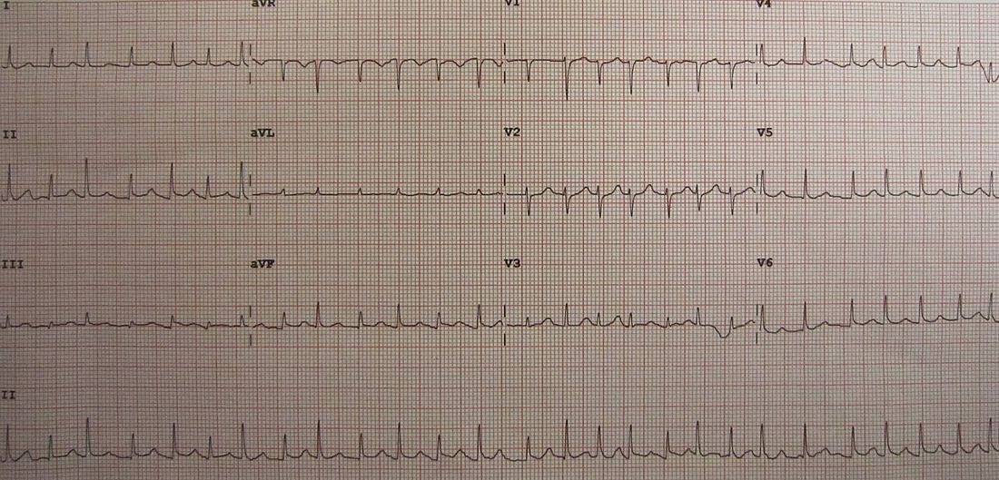
Pulsus paradoxus is a drop in systolic pressure of 10 mm Hg or more on inspiration during normal breathing. It also occurs in acute asthma, massive pulmonary embolism, constrictive pericarditis and hypovolemia.10
Bedside echo
Effusion size by the ESC, measured as the largest echo-free space between the heart and the pericardium at end-diastole.1 A large effusion (over 20 mm) is a high-risk feature in pericarditis.1
- Emergency physicians can find the fluid. In 515 high-risk ED patients, emergency physician echo detected effusions with 96% sensitivity and 98% specificity against cardiology review.5
- Diastolic right ventricular collapse is the specific sign. Systolic right atrial collapse and a plethoric, non-collapsing IVC are the sensitive ones.3
- Sonographic pulsus paradoxus is another sign.3
- No sign is universal. In 239 ED patients drained within 48 hours, 69.1% had RV diastolic collapse, 62.3% had exaggerated mitral inflow variation, 56.4% had a plethoric IVC, and 53.4% had RA systolic collapse.11
ESC triage score
The ESC uses a triage score, built by expert consensus, to decide whether to drain at once or delay.1,9 Hemodynamic instability, impending deterioration or cardiac arrest call for emergent drainage.3
| Category | Item | Points |
|---|---|---|
| Cause | ||
| Cause | Malignant disease; tuberculosis | 2 each |
| Cause | Recent radiotherapy; recent viral infection; recurrent effusion; end-stage renal failure; immunosuppression | 1 each |
| Cause | Thyroid disease; systemic autoimmune disease | −1 each |
| Clinical picture | ||
| Clinical | Orthopnea | 3 |
| Clinical | Pulsus paradoxus > 10 mm Hg; rapid worsening | 2 each |
| Clinical | Dyspnea or tachypnea; progressive sinus tachycardia; oliguria | 1 each |
| Clinical | Hypotension (central BP < 95 mm Hg); pericardial chest pain; friction rub | 0.5 each |
| Clinical | Slow course | −1 |
| Imaging | ||
| Imaging | Large circumferential effusion | 3 |
| Imaging | Left atrial collapse | 2 |
| Imaging | Plethoric non-collapsing IVC; right ventricular collapse | 1.5 each |
| Imaging | Moderate effusion; right atrial collapse; mitral/tricuspid respiratory flow variation; swinging heart; cardiomegaly on X-ray; low voltage on ECG | 1 each |
| Imaging | Electrical alternans | 0.5 |
| Imaging | Small effusion | −1 |
Restated from the 2025 ESC guideline (Figure 12), which adopted the 2014 position statement of its working group.1,9 The guideline's text and caption say more than 6 points from at least two categories means urgent drainage. Its flowchart box says 6 or more. Below that, the flowchart postpones drainage and starts empiric anti-inflammatory therapy. After drainage, patients who improve go to outpatient follow-up, and those who don't get a pericardial window.1
Before the needle
- Avoid positive-pressure ventilation and IV sedation if you can. Both can lower cardiac output.3
- Fluid is a bridge, not a treatment. In 49 patients with hemodynamic tamponade given 500 mL saline over 10 minutes, cardiac index rose by more than 10% in 47%, was unchanged in 22%, and fell in 31%. A systolic pressure under 100 mm Hg predicted a good response.7
- Give blood for traumatic hemopericardium. Treat anticoagulation, coagulopathy and anemia.3
- Consider vasoactive drugs in hypotensive patients, alongside gentle fluids.3 Drainage is the treatment.2
Drainage
- Pericardiocentesis (echo-, CT- or fluoroscopy-guided) is recommended for tamponade, suspected bacterial or cancer-related pericarditis, and symptomatic moderate to large effusions despite medical therapy (Class I, level C).1
- Echo guidance works. In 1,127 echo-guided pericardiocenteses at one center, the success rate was 97%. Complications occurred in 4.7% (major 1.2%).8
- Technique: a 16–20 gauge needle, usually from below the xiphoid; an apical or left lateral approach is possible. Pick the largest, shallowest pocket with nothing vital in the way.1,3
- Confirm the needle is in the pericardium with agitated saline and a look at the first aspirate for blood.3
- Drain slowly. Take usually under 500 mL at first to avoid pericardial decompression syndrome. Leave a drain in until output falls under 30 mL a day.1,3
When to go to surgery instead
- Type A aortic dissection with hemopericardium, ventricular free wall rupture after MI, severe chest trauma, and procedural bleeding that can't be controlled through a catheter.2,3
- Purulent effusions, and effusions a needle can't reach (Class I, level C).1
- Effusions that keep coming back despite medical therapy: a pleuro-pericardial window (Class I, level C).1
Causes
- Common, most frequent first: cancer, procedures and trauma, pericarditis, and tuberculosis (the most common cause in low-income countries).1
- Less common: autoimmune disease, pericardial injury syndromes, acute MI, aortic dissection, uremia, bacterial infection, and pneumopericardium.1
- Procedures are now a leading cause. In the Mayo Clinic series, cardiac surgery, cancer and catheter-procedure perforations together made up nearly 70% of drainages.8
- Acute pericarditis rarely causes tamponade: under 1% of cases.1
Disposition
- Tamponade: admit. Signs of tamponade and a large effusion are high-risk features in the ESC pericarditis risk table, and the ESC recommends admitting high-risk patients.1
- Effusion without tamponade: the ESC score helps decide whether drainage can be delayed.1 Drainage is also indicated when the cause must be found, as in suspected cancer or bacterial infection.1
- After drainage: NSAIDs and colchicine can be considered to prevent recurrence.2
Pitfalls
- Waiting for hypotension or the full Beck triad.3,7
- Intubating before drainage without planning for the drop in cardiac output.3
- Assuming a big fluid bolus will fix it. It helped only about half of patients in one study.7
- Sending a patient with chest trauma or aortic dissection for needle drainage instead of surgery.2,3
- Draining a large effusion all at once.1
- Reading a pulsus paradoxus as tamponade without thinking of asthma, massive PE or hypovolemia.10
How strong is the evidence?
On-shift teaching
Questions to ask a learner at the bedside. Tap a question to see the answer. Every answer uses facts cited above.
Q1A patient has a moderate effusion. How much does pulsus paradoxus change your estimate of tamponade?
Q2Which echo signs are specific for tamponade, and which are sensitive?
Q3How reliable is emergency physician echo for finding an effusion?
Q4Why avoid intubation in tamponade if you can?
Q5Does a fluid bolus help?
References
What this page is built on
11 sources, cited 106 times. Dots are sources, filled by how far each was read; stronger color means a larger share of the citations. Tap a tier to see its sources.
Checked by the writer, not a physician. The dots show how each source was read, not that anyone has confirmed the claims.
- 1Schulz-Menger J, Collini V, Gröschel J, et al. 2025 ESC Guidelines for the management of myocarditis and pericarditis. Eur Heart J. 2025;46(40):3952–4041.
- 2Adler Y, Ristić AD, Imazio M, et al. Cardiac tamponade. Nat Rev Dis Primers. 2023;9(1):36.
- 3Alerhand S, Adrian RJ, Long B, Avila J. Pericardial tamponade: a comprehensive emergency medicine and echocardiography review. Am J Emerg Med. 2022;58:159–174.
- 4Roy CL, Minor MA, Brookhart MA, Choudhry NK. Does this patient with a pericardial effusion have cardiac tamponade? JAMA. 2007;297(16):1810–1818.
- 5Mandavia DP, Hoffner RJ, Mahaney K, Henderson SO. Bedside echocardiography by emergency physicians. Ann Emerg Med. 2001;38(4):377–382.
- 6Mathur AP, Saini A, Lucas BP, et al. Diagnostic accuracy retrospectively of electrocardiographic findings and cancer history for tamponade in patients determined to have pericardial effusion by transthoracic echocardiogram. Am J Cardiol. 2013;111(7):1062–1066.
- 7Sagristà-Sauleda J, Angel J, Sambola A, Permanyer-Miralda G. Hemodynamic effects of volume expansion in patients with cardiac tamponade. Circulation. 2008;117(12):1545–1549.
- 8Tsang TS, Enriquez-Sarano M, Freeman WK, et al. Consecutive 1127 therapeutic echocardiographically guided pericardiocenteses: clinical profile, practice patterns, and outcomes spanning 21 years. Mayo Clin Proc. 2002;77(5):429–436.
- 9Ristić AD, Imazio M, Adler Y, et al. Triage strategy for urgent management of cardiac tamponade: a position statement of the European Society of Cardiology Working Group on Myocardial and Pericardial Diseases. Eur Heart J. 2014;35(34):2279–2284.
- 10Life in the Fast Lane. Pulsus paradoxus.
- 11Eke OF, Jalbout NA, Selame L, et al. Pericardial tamponade: a new perspective on echocardiographic features and application of a prediction score. Intern Emerg Med. 2024;19(6):1757–1764.
AI-generated draft. Not reviewed by a physician. This page is for education only and may contain errors, including in doses. Do not use it to diagnose, treat or make decisions about any patient. Rely on your own clinical judgment, your institution's protocols and current drug labeling. Reading this page does not create a physician-patient relationship. Full terms