Acute kidney injury
Bottom line
- Define it by change, not by a single value. AKI is a creatinine rise of ≥ 0.3 mg/dL within 48 hours, a rise to ≥ 1.5 times baseline within 7 days, or urine output < 0.5 mL/kg/h for 6 hours.2 Compare every creatinine with a baseline.1
- First ask whether the patient needs dialysis now. Hyperkalemia, acidosis, fluid overload, pulmonary edema or uremic complications that don't respond to treatment need immediate referral.1
- Restore volume with isotonic crystalloid, and stop the harm. Crystalloid is suggested over colloid.2 Review NSAIDs, aminoglycosides, ACE inhibitors, ARBs and diuretics.1
- Dipstick every patient's urine; image selectively. Blood plus protein without infection suggests nephritis. Ultrasound within 6 hours for a suspected infected, obstructed kidney, within 24 hours when no cause is found.1
- Don't treat AKI with diuretics or dopamine, and don't fear needed contrast. Neither drug helps the kidney.1,2 In ED cohorts, IV contrast for CT was not linked to more AKI.9,10
Definition and stages
| Stage | Serum creatinine | Urine output |
|---|---|---|
| 1 | 1.5–1.9 × baseline, or a rise of ≥ 0.3 mg/dL (≥ 26.5 µmol/L) | < 0.5 mL/kg/h for 6–12 h |
| 2 | 2.0–2.9 × baseline | < 0.5 mL/kg/h for ≥ 12 h |
| 3 | ≥ 3.0 × baseline, or a rise to ≥ 4.0 mg/dL (≥ 353.6 µmol/L), or start of dialysis | < 0.3 mL/kg/h for ≥ 24 h, or no urine for ≥ 12 h |
KDIGO staging. Use the worse of the two columns.2 NICE uses the same definitions and gives the absolute rise as 26 µmol/L within 48 hours.1 For creatinine, 1 mg/dL is 88.4 µmol/L.
- Who to test: acutely ill adults with chronic kidney disease (especially eGFR < 60), heart failure, liver disease, diabetes, prior AKI, low urine output, hypovolemia, sepsis, a worsening early warning score, age ≥ 65, or limited access to fluids because they rely on a carer.1
- Recent exposures that count: NSAIDs, aminoglycosides, ACE inhibitors, ARBs or diuretics in the past week (especially with hypovolemia), iodinated contrast in the past week, and known or likely urinary obstruction.1
- In chronic kidney disease, a creatinine rise may be AKI rather than slow progression.1
- Why it matters in the ED: in a UK prospective cohort, AKI was found in 2.8% of adult ED attendances. In-patient mortality was 24.4%, against 3.2% without AKI, and 38.8% when the stage worsened after admission.3
First steps
- Look for a dialysis emergency. Hyperkalemia, metabolic acidosis, fluid overload, pulmonary edema, or uremic complications such as pericarditis or encephalopathy that don't respond to medical treatment need immediate referral for renal replacement therapy.1
- Assess volume and perfusion. Give isotonic crystalloid for volume expansion rather than albumin or starch, unless there is hemorrhagic shock. Add vasopressors to fluids in vasodilatory shock.2
- Stop or adjust harmful drugs. Consider holding ACE inhibitors and ARBs during vomiting, diarrhea or sepsis until the patient is stable. Ask a pharmacist to review doses.1
- Dipstick the urine for blood, protein, leukocytes, nitrites and glucose as soon as AKI is suspected.1
- Decide on ultrasound by the likely cause (see below).1
- Recheck creatinine and record urine output. Daily creatinine is typical in hospital; monitor more often if the patient needs it.1,2
- At-risk adult, acutely ill. Measure creatinine and compare it with a baseline.1
- Confirm and stage: rise ≥ 0.3 mg/dL in 48 h, ≥ 1.5 × baseline within 7 days, or urine < 0.5 mL/kg/h for 6 h.2
- No, or while you wait
- Either wayStop the harm. Review NSAIDs, aminoglycosides, ACE inhibitors, ARBs and diuretics. Consider holding ACE inhibitors and ARBs in vomiting, diarrhea or sepsis.1
- Dipstick the urine. Blood and protein without infection or catheter trauma: think nephritis and involve nephrology.1
- Suspected infected, obstructed kidney?YesUltrasound within 6 h and refer to urology now. Decompress with nephrostomy or stent within 12 h.1
- NoNo cause found, or obstruction possible: ultrasound within 24 h. Skip routine ultrasound when the cause is clear.1
- Contrast CT needed? Don't delay it in an emergency.1
- Talk to nephrology within 24 h for stage 3, no clear cause, poor response, a transplant, CKD stage 4–5, or possible vasculitis, glomerulonephritis or myeloma. Recheck creatinine.1
Finding the cause
- Find and record a cause in every patient, with attention to the reversible ones.1,2
- Urine dipstick: hematuria and proteinuria with no obvious cause, and without a urinary infection or catheter trauma, should raise acute nephritis and prompt nephrology referral.1
- Specialist diagnoses to consider: vasculitis, glomerulonephritis, tubulointerstitial nephritis and myeloma.1
- Multi-system disease: think of it when AKI comes with signs elsewhere, such as a purpuric rash.1
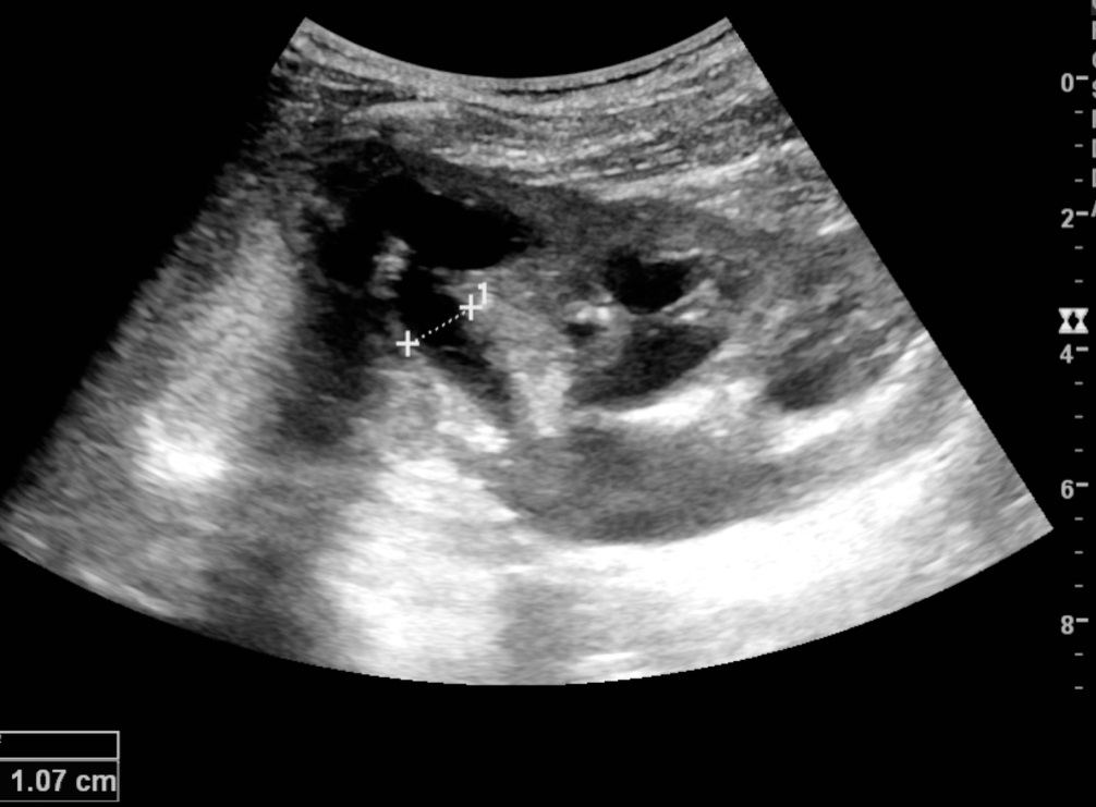
Ultrasound
| Situation | Ultrasound |
|---|---|
| Suspected pyonephrosis (infected and obstructed kidney) | Immediately, within 6 hours1 |
| No cause identified, or at risk of urinary obstruction | Urgently, within 24 hours1 |
| Cause already identified | Not routinely1 |
In 797 inpatients with AKI who had an ultrasound, 10.6% had hydronephrosis and 3.3% needed an intervention for it. In the low-risk group, only 1 of 223 (0.4%) needed an intervention. Prior hydronephrosis, recurrent urinary infections and a clinical picture of obstruction pointed to higher risk; nephrotoxic drugs, heart failure or a prerenal cause pointed to lower risk.4
Treatment
| Measure | Recommendation | Source and strength |
|---|---|---|
| Use | ||
| Isotonic crystalloid | Initial volume expansion, rather than albumin or starch, unless there is hemorrhagic shock | KDIGO, 2B2 |
| Vasopressors | With fluids in vasodilatory shock | KDIGO, 1C2 |
| Loop diuretics | Only for fluid overload or edema, while awaiting dialysis or during recovery | NICE; KDIGO, 2C1,2 |
| Avoid | ||
| Loop diuretics to treat AKI itself | Not routinely | NICE; KDIGO, 2C1,2 |
| Low-dose dopamine | Do not use to prevent or treat AKI | NICE; KDIGO, 1A1,2 |
| Aminoglycosides | Avoid unless no suitable, less nephrotoxic alternative exists | KDIGO, 2A2 |
KDIGO grades: 1 is a recommendation and 2 a suggestion; A to D is the quality of evidence, from high to very low.2 No drug doses are given here; none is specific to AKI.
Balanced crystalloid or saline?
- SMART (15,802 critically ill adults): balanced crystalloids lowered the 30-day composite of death, new dialysis or persistent kidney dysfunction, 14.3% vs 15.4%.5
- SALT-ED (13,347 non-critically ill ED patients, median 1,079 mL in the ED): hospital-free days were the same, but the same kidney composite was lower with balanced fluids, 4.7% vs 5.6%.6
Dialysis
- Indications: refer immediately when hyperkalemia, metabolic acidosis, uremic complications, fluid overload or pulmonary edema fail medical treatment. Start dialysis emergently for life-threatening fluid, electrolyte or acid-base changes.1,2
- Judge the whole patient, not an isolated urea, creatinine or potassium value.1,2
- Starting early doesn't help. In STARRT-AKI (2,927 critically ill adults), starting within 12 hours of eligibility did not lower 90-day mortality (43.9% vs 43.7%). Survivors were more often still dialysis-dependent (10.4% vs 6.0%), and adverse events were more common (23.0% vs 16.5%).7
Contrast and the kidney
- Don't delay emergency contrast when the delay itself is likely to cause harm.1 For patients on dialysis or with a transplant, NICE advises talking to nephrology first, but not delaying emergency imaging for it.1
- The risk is small. NICE notes a small but increased risk of AKI when eGFR is below 30.1 The American College of Radiology and the National Kidney Foundation say the risk has been overstated, mainly because older studies lacked control groups.8
- Meta-analysis: across 28 observational studies (107,335 patients), contrast CT was not linked to AKI (OR 0.94, 0.83–1.07), dialysis (OR 0.83, 0.59–1.16) or death (OR 1.0, 0.73–1.36) compared with non-contrast CT.9,13
- ED cohort: in 17,934 ED visits, contrast was not linked to AKI by KDIGO criteria (OR 1.00, 0.87–1.16), whatever the baseline kidney function.10
- Patients who already have AKI: in 14,449 ED patients arriving with AKI, contrast was not linked to persistent AKI at discharge (OR 0.93, 0.83–1.05) or to dialysis within 180 days.11
- Prophylaxis: in PRESERVE (4,993 high-risk patients having angiography), IV bicarbonate was no better than IV saline, and oral acetylcysteine no better than placebo.12
On IV fluids before contrast: KDIGO 2012 recommends IV saline or bicarbonate over no IV fluid for anyone at increased risk (1A), and suggests adding oral acetylcysteine (2D).2 The American College of Radiology and the National Kidney Foundation reserve IV saline for patients with AKI or eGFR < 30 who are not on dialysis.8 NICE suggests only considering it for inpatients at particularly high risk, such as eGFR < 30 or a transplant.1 PRESERVE, published after KDIGO, found no benefit from acetylcysteine.12
Referral and follow-up
- Immediately: anyone who meets the dialysis criteria above.1
- Within 24 hours: a possible specialist diagnosis (vasculitis, glomerulonephritis, tubulointerstitial nephritis, myeloma), no clear cause, a poor response to treatment, complications, stage 3, a kidney transplant, or CKD stage 4–5.1
- Nephrology not needed: a clear cause that is responding promptly to treatment, unless the patient has a transplant.1
- After the episode: keep checking creatinine, and reassess for new or worse chronic kidney disease at 3 months.1,2 Talk with patients about the risks of dehydration and of over-the-counter NSAIDs.1
Pitfalls
- Calling a creatinine "normal" without a baseline. A rise of 0.3 mg/dL in 48 hours is already AKI.2
- Giving furosemide to make urine. Loop diuretics don't treat AKI; use them only for fluid overload.1,2
- Using low-dose dopamine "for the kidneys".1,2
- Withholding a contrast CT that the patient needs now.1,9,10
- Starting dialysis for a number alone. Decide on the whole patient; starting early did not improve survival.1,7
- Leaving ACE inhibitors, ARBs and NSAIDs on the medication list of a vomiting or septic patient.1
- Missing an infected, obstructed kidney. It needs ultrasound within 6 hours and decompression within 12.1
How strong is the evidence?
The definition and staging are expert consensus and not graded.2 Most KDIGO treatment statements are suggestions (grade 2), with the firmest evidence against low-dose dopamine (1A).2 Most NICE AKI recommendations date from 2013.1 The fluid trials are large and randomized.5,6 The contrast data in CT are observational, because randomizing patients to no contrast is hard to do.9,13
On-shift teaching
Questions to ask a learner at the bedside. Tap a question to see the answer. Every answer uses facts cited above.
Q1What are the three ways a patient can meet the KDIGO definition of AKI?
Q2What puts a patient in stage 3?
Q3Which complications mean you should refer for dialysis immediately?
Q4Did starting dialysis early help in STARRT-AKI?
Q5Which patients with AKI need an ultrasound within 6 hours?
References
What this page is built on
14 sources, cited 105 times. Dots are sources, filled by how far each was read; stronger color means a larger share of the citations. Tap a tier to see its sources.
Checked by the writer, not a physician. The dots show how each source was read, not that anyone has confirmed the claims.
- 1National Institute for Health and Care Excellence. Acute kidney injury: prevention, detection and management (NG148). Published December 2019; last updated October 2024.
- 2Kidney Disease: Improving Global Outcomes (KDIGO) Acute Kidney Injury Work Group. KDIGO clinical practice guideline for acute kidney injury. Kidney Int Suppl. 2012;2(1):1–138.
- 3Foxwell DA, Pradhan S, Zouwail S, et al. Epidemiology of emergency department acute kidney injury. Nephrology (Carlton). 2020;25(6):457–466.
- 4Licurse A, Kim MC, Dziura J, et al. Renal ultrasonography in the evaluation of acute kidney injury: developing a risk stratification framework. Arch Intern Med. 2010;170(21):1900–1907.
- 5Semler MW, Self WH, Wanderer JP, et al. Balanced crystalloids versus saline in critically ill adults (SMART). N Engl J Med. 2018;378(9):829–839.
- 6Self WH, Semler MW, Wanderer JP, et al. Balanced crystalloids versus saline in noncritically ill adults (SALT-ED). N Engl J Med. 2018;378(9):819–828.
- 7STARRT-AKI Investigators; Bagshaw SM, Wald R, Adhikari NKJ, et al. Timing of initiation of renal-replacement therapy in acute kidney injury. N Engl J Med. 2020;383(3):240–251.
- 8Davenport MS, Perazella MA, Yee J, et al. Use of intravenous iodinated contrast media in patients with kidney disease: consensus statements from the American College of Radiology and the National Kidney Foundation. Kidney Med. 2020;2(1):85–93.
- 9Aycock RD, Westafer LM, Boxen JL, et al. Acute kidney injury after computed tomography: a meta-analysis. Ann Emerg Med. 2018;71(1):44–53.e4.
- 10Hinson JS, Ehmann MR, Fine DM, et al. Risk of acute kidney injury after intravenous contrast media administration. Ann Emerg Med. 2017;69(5):577–586.e4.
- 11Ehmann MR, Mitchell J, Levin S, et al. Renal outcomes following intravenous contrast administration in patients with acute kidney injury: a multi-site retrospective propensity-adjusted analysis. Intensive Care Med. 2023;49(2):205–215.
- 12Weisbord SD, Gallagher M, Jneid H, et al. Outcomes after angiography with sodium bicarbonate and acetylcysteine (PRESERVE). N Engl J Med. 2018;378(7):603–614.
- 13Rezaie S. Is contrast induced nephropathy (CIN) really not a thing? REBEL EM; September 25, 2017.
- 14Emergency Medicine Cases. Acute kidney injury. Further reading; not used for any claim above.
AI-generated draft. Not reviewed by a physician. This page is for education only and may contain errors, including in doses. Do not use it to diagnose, treat or make decisions about any patient. Rely on your own clinical judgment, your institution's protocols and current drug labeling. Reading this page does not create a physician-patient relationship. Full terms